
Page 206 - An Introduction to Microelectromechanical Systems Engineering
P. 206
Summary 185
method is sufficiently sensitive to detect single base differences and single-point
mutations in the DNA sequence.
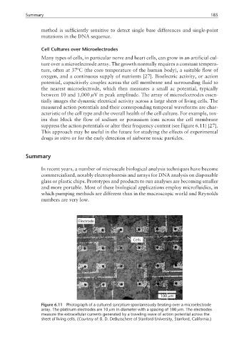
Cell Cultures over Microelectrodes
Many types of cells, in particular nerve and heart cells, can grow in an artificial cul-
ture over a microelectrode array. The growth normally requires a constant tempera-
ture, often at 37ºC (the core temperature of the human body), a suitable flow of
oxygen, and a continuous supply of nutrients [27]. Bioelectric activity, or action
potential, capacitively couples across the cell membrane and surrounding fluid to
the nearest microelectrode, which then measures a small ac potential, typically
between 10 and 1,000 µV in peak amplitude. The array of microelectrodes essen-
tially images the dynamic electrical activity across a large sheet of living cells. The
measured action potentials and their corresponding temporal waveforms are char-
acteristic of the cell type and the overall health of the cell culture. For example, tox-
ins that block the flow of sodium or potassium ions across the cell membrane
suppress the action potentials or alter their frequency content (see Figure 6.11) [27].
This approach may be useful in the future for studying the effects of experimental
drugs in vitro or for the early detection of airborne toxic particles.
Summary
In recent years, a number of microscale biological analysis techniques have become
commercialized, notably electrophoresis and arrays for DNA analysis on disposable
glass or plastic chips. Prototypes and products to run analyses are becoming smaller
and more portable. Most of these biological applications employ microfluidics, in
which pumping methods are different than in the macroscopic world and Reynolds
numbers are very low.
Electrode
Cells
µ
100 m
Figure 6.11 Photograph of a cultured syncytium spontaneously beating over a microelectrode
array. The platinum electrodes are 10 µm in diameter with a spacing of 100 µm. The electrodes
measure the extracellular currents generated by a traveling wave of action potential across the
sheet of living cells. (Courtesy of: B. D. DeBusschere of Stanford University, Stanford, California.)