
Page 236 - Artificial Intelligence in the Age of Neural Networks and Brain Computing
P. 236
4. Electrophysiological Time-Series 227
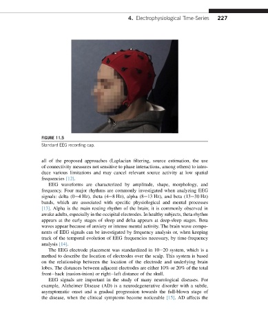
FIGURE 11.5
Standard EEG recording cap.
all of the proposed approaches (Laplacian filtering, source estimation, the use
of connectivity measures not sensitive to phase interactions, among others) to intro-
duce various limitations and may cancel relevant source activity at low spatial
frequencies [12].
EEG waveforms are characterized by amplitude, shape, morphology, and
frequency. Four major rhythms are commonly investigated when analyzing EEG
signals: delta (0e4 Hz), theta (4e8 Hz), alpha (8e13 Hz), and beta (13e30 Hz)
bands, which are associated with specific physiological and mental processes
[13]. Alpha is the main resting rhythm of the brain; it is commonly observed in
awake adults, especially in the occipital electrodes. In healthy subjects, theta rhythm
appears at the early stages of sleep and delta appears at deep-sleep stages. Beta
waves appear because of anxiety or intense mental activity. The brain wave compo-
nents of EEG signals can be investigated by frequency analysis or, when keeping
track of the temporal evolution of EEG frequencies necessary, by time-frequency
analysis [14].
The EEG electrode placement was standardized in 10e20 system, which is a
method to describe the location of electrodes over the scalp. This system is based
on the relationship between the location of the electrode and underlying brain
lobes. The distances between adjacent electrodes are either 10% or 20% of the total
fronteback (nasion-inion) or righteleft distance of the skull.
EEG signals are important in the study of many neurological diseases. For
example, Alzheimer Disease (AD) is a neurodegenerative disorder with a subtle,
asymptomatic onset and a gradual progression towards the full-blown stage of
the disease, when the clinical symptoms become noticeable [15]. AD affects the