
Page 133 - Biomedical Engineering and Design Handbook Volume 1, Fundamentals
P. 133
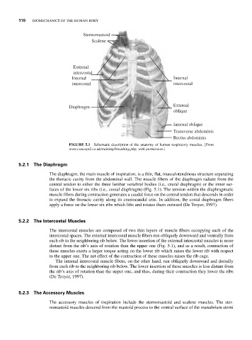
110  BIOMECHANICS OF THE HUMAN BODY

                                    Sternomastoid
                                       Scalene


                                External
                                intercostal
                               Internal                                Internal
                               intercostal                             intercostal


                              Diaphragm                                External
                                                                       oblique

                                                                       Internal oblique
                                                                       Transverse abdominis
                                                                       Rectus abdominis
                              FIGURE 5.1  Schematic description of the anatomy of human respiratory muscles. [From
                              www.concept2.co.uk/training/breathing.php, with permission.]


           5.2.1 The Diaphragm
                       The diaphragm, the main muscle of inspiration, is a thin, flat, musculotendinous structure separating
                       the thoracic cavity from the abdominal wall. The muscle fibers of the diaphragm radiate from the
                       central tendon to either the three lumbar vertebral bodies (i.e., crural diaphragm) or the inner sur-
                       faces of the lower six ribs (i.e., costal diaphragm) (Fig. 5.1). The tension within the diaphragmatic
                       muscle fibers during contraction generates a caudal force on the central tendon that descends in order
                       to expand the thoracic cavity along its craniocaudal axis. In addition, the costal diaphragm fibers
                       apply a force on the lower six ribs which lifts and rotates them outward (De Troyer, 1997).


           5.2.2 The Intercostal Muscles
                       The intercostal muscles are composed of two thin layers of muscle fibers occupying each of the
                       intercostal spaces. The external intercostal muscle fibers run obliquely downward and ventrally from
                       each rib to the neighboring rib below. The lower insertion of the external intercostal muscles is more
                       distant from the rib’s axis of rotation than the upper one (Fig. 5.1), and as a result, contraction of
                       these muscles exerts a larger torque acting on the lower rib which raises the lower rib with respect
                       to the upper one. The net effect of the contraction of these muscles raises the rib cage.
                         The internal intercostal muscle fibers, on the other hand, run obliquely downward and dorsally
                       from each rib to the neighboring rib below. The lower insertion of these muscles is less distant from
                       the rib’s axis of rotation than the upper one, and thus, during their contraction they lower the ribs
                       (De Troyer, 1997).


           5.2.3 The Accessory Muscles
                       The accessory muscles of inspiration include the sternomastoid and scalene muscles.  The ster-
                       nomastoid muscles descend from the mastoid process to the ventral surface of the manubrium sterni