
Page 141 - Biomedical Engineering and Design Handbook Volume 1, Fundamentals
P. 141
118 BIOMECHANICS OF THE HUMAN BODY
Rib cage
muscles
Bony skeleton
other than
Ppl L rib cage
RC pul Central
tendon
Spring
RC ab Pab Crural
Ppl ab diaphragm
Crural
diaphragm
Anterior
abdominal
wall
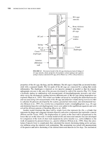
FIGURE 5.5 Mechanical model of the rib cage showing mechanical linkage of
rib cage muscles, elastic properties of respiratory system (springs) and agencies
acting to displace and distort rib cage. [From Ward et al. (1992), with permission.]
properties of the rib cage, the lung, and the abdomen. The rib cage is shaped like an inverted hockey
stick with a separated handle. The two parts of the rib cage are connected by a spring that resists
deformation. The diaphragm is depicted as two muscles arranged in parallel so that the transdi-
aphragmatic pressure is the sum of the pressure developed by each of the muscles (Fig. 5.5). Using
a hydraulic analog in combination with measurements of transdiaphragmatic pressures and relax-
ation curves the mechanical coupling between different parts of the rib cage during inspiration was
explored (Ward et al., 1992). This model was further advanced by including the abdominal muscles
and was used along with measurements of the rib cage and abdomen volume during exercise in order
to calculate the pressure developed by the scalene, parasternal intercostals, and sternomastoid mus-
cles (Kenyon et al., 1997). In a similar two-compartment model, extradiaphragmatic (e.g., rib cage
and abdominal muscles) and diaphragmatic forces were added in the equilibrium equations and were
solved for different patterns of breathing (Ricci et al., 2002).
Another model simulated the chest wall by simple levers that represent the ribs, a cylinder that
represents the lungs and a diagonal element of passive and active components that represents a muscle
(Wilson and De Troyer, 1992). The displacement of a point on the chest wall is proportional to the
forces that act on the chest wall. A similar model of ribs and intercostal muscles was also developed
for comparison of the work of chest wall expansion by active muscles (i.e., active inflation) to the
work of expansion by pressure forces (i.e., passive inflation) (Wilson et al., 1999). Since the calcu-
lation of muscle force is complicated, they calculated the muscle shortening during active and pas-
sive inflation using the minimal work assumption. This assumption was tested with measurements
of the passive and active shortening of the internal intercostal muscles in five dogs. The mechanical