Page 142 - Biomedical Engineering and Design Handbook Volume 1, Fundamentals
P. 142

BIOMECHANICS OF THE RESPIRATORY MUSCLES  119

                          analogs of all the models described so far have not differentiated between individual groups of respi-
                          ratory muscles and between the rib cage and the accessory muscles, and thus precluded evaluation
                          of their contribution to respiration.
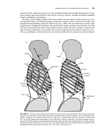
                            Recently, a more realistic model of the chest wall has been developed with the objective to eval-
                          uate the performance of individual groups of respiratory muscles in terms of the force and the work
                          generated during breathing maneuvers (Ratnovsky et al., 2005). The two-dimensional model in the
                          sagittal plane is composed of stationary and mobile bones and the respiratory muscles (Fig. 5.6). The
                          stationary elements are the bones of the vertebral column, skull, iliac crest, and pelvis, while the rib
                          cage forms the mobile part of the model. The model incorporates three groups of inspiratory mus-
                          cles (e.g., diaphragm, external intercostal, and sternomastoid) and four groups of expiratory muscles


                               A                               B


                              Hinge
                                            SM
                                                             Skull
                                                                   γ i
                                                                       α i
                                                                                    Rib
                              L T1         EI 1
                                                         Sternum
                                                                                      Lung

                                                                                        Central
                                                                                        tendon


                                                                                   CD 1
                         Vertebral                      EO 1
                                                                            CD 4
                           column
                                                       EO 2
                                      EI 23                                     CD 2
                                                                              CD 3
                                                     EO 3           RD
                                           EI 24
                                                                       TR          RC
                           Iliac crest             EO 4
                                               EO 6                      IO               Abdominal
                                            EO 8
                                                                                          wall


                          FIGURE 5.6 Scheme of a two-dimensional model of the human trunk in the sagittal plane. The springs represent the
                          respiratory muscles while the thick lines represent the vertebral column, sternum, and iliac crest. α represents the rib
                                                                                      i
                          angles, γ represents the vertebral column curvature angles, and L represents the length of each thorax vertebra. SM,
                               i                                  Ti
                          RC, TR, and IO represent the sternomastoid, rectus abdominis, transverse abdominis, and internal oblique muscles,
                          respectively. EI -EI represent the external intercostal muscle units and their angle with the horizon. EO -EO represent
                                   1  24                                                1  6
                          the external oblique muscle units. CD , CD , CD , CD , and RD represent the costal fibers and the crural fibers of the
                                                 1  2   3  4
                          diaphragm muscle, respectively. [From Ratnovsky et al. (2005), with permission.]
   137   138   139   140   141   142   143   144   145   146   147