
Page 389 - Biomedical Engineering and Design Handbook Volume 2, Applications
P. 389
BREAST IMAGING SYSTEMS: DESIGN CHALLENGES FOR ENGINEERS 367
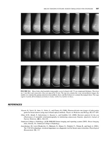
FIGURE 12.6 Slices from a dual modality tomographic scan of a breast with 7.9 cm compressed thickness. The slices
are 1 mm thick and consecutive slices are spaced by 10 mm. The top row contains the x-ray tomosynthesis images; the
middle row contains the gamma emission tomosynthesis slices; and the bottom row contains the merged slices. The large
region of poorly differentiated carcinoma is clearly visible in a number of slices.
REFERENCES
Alacam, B., Yazici, B., Intes, X., Nioka, S., and Chance, B. (2008). Pharmacokinetic-rate images of indocyanine
green for breast tumors using near-infrared optical methods. Physics in Medicine and Biology. 53, 837–859.
Allen, M.W., Hendi, P., Schwimmer, J., Bassett, L., and Gambhir, S.S. (2000). Decision analysis for the cost
effectiveness of sestamibi scintimammography in minimizing unnecessary biopsies. Quarterly Journal of
Nuclear Medicine. 44(2), 168–185.
American College of Radiology (ACR) BIRADS breast imaging and reporting system (2003). Breast Imaging
Atlas, Reston, VA: American College of Radiology.
Assenheimer, M., Laver-Moskovitz, O., Malonek, D., Manor, D., Nahaliel, U., Nitzan, R., and Saad, A. (2001).
The T-SCAN technology: electrical impedance as a diagnostic tool for breast cancer detection. Physiological
Measurement. 22, 1–8.