
Page 77 - Carbon Nanotubes
P. 77
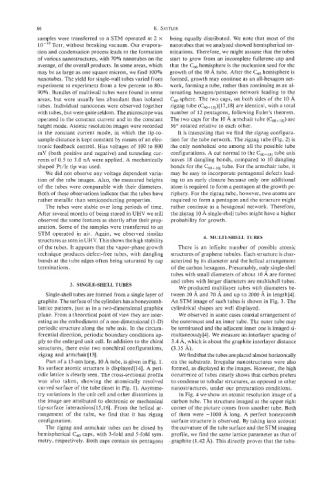
66                                    K.  SATTLER
             samples were transferred  to a STM operated at 2 x   being equally distributed.  We note that most of the
             lo-''  Torr, without breaking vacuum. Our evapora-  nanotubes that we analyzed showed hemispherical ter-
             tion and condensation process leads to the formation   minations. Therefore, we might assume that the tubes
             of various nanostructures, with 70% nanotubes on the   start to grow from an incomplete fullerene cap and
             average, of the overall products. In some areas, which  that the C,,  hemisphere is the nucleation seed for the
             may be as large as one square micron, we find 100%  growth of the 10 A tube. After the C6'  hemisphere is
             nanotubes. The yield for single-wall tubes varied from  formed, growth may continue as an all-hexagon net-
             experiment to experiment from a few percent to 80-   work, forming a tube, rather than continuing as an al-
             90%. Bundles of multiwall tubes were found in some  ternating hexagodpentagon network  leading to the
             areas,  but were usually less abundant than isolated   C6, sphere. The two caps, on both sides of the 10 A
             tubes.  Individual nanocones were observed together   zigzag tube (C60+18j)[17,1S] are identical, with a total
             with tubes, but were quite seldom. The microscope was   number of  12 pentagons, following Euler's theorem.
             operated in the constant current and in the constant   The two caps for the 10 A armchair tube (C60+10j) are
             height mode. Atomic resolution images were recorded   36" rotated  relative to each other.
             in  the  constant  current  mode,  in  which  the tip-to-   It is interesting that we find the zigzag configura-
             sample distance is kept constant by means of an elec-  tion for the tube network. The zigzag tube (Fig. 2) is
             tronic feedback control.  Bias voltages of  100 to 800   the only nonhelical one among all the possible tube
             mV  (both positive and negative) and tunneling  cur-  configurations.  A cut normal to the C60+18j tube axis
             rents of 0.5 to 3.0 nA were applied. A mechanically   leaves  18 dangling bonds,  compared to 10 dangling
             shaped Pt/lr  tip was used.                 bonds for the C60+10j tube. For the armchair tube, it
                We did not observe any voltage dependent varia-   may be easy to incorporate pentagonal defects lead-
             tion of the tube images. Also, the measured heights   ing to an early closure because only one additional
              of  the tubes  were comparable with their  diameters.   atom is required to form a pentagon at the growth pe-
              Both of these observations indicate that the tubes have   riphery. For the zigzag tube, however, two atoms are
              rather metallic than semiconducting properties.   required to form a pentagon and the structure might
                The tubes were stable over long periods of time.   rather  continue as a hexagonal network. Therefore,
              After several months of being stored in UHV we still   the zigzag 10 A single-shell tubes might have a higher
              observed the same features as shortly after their prep-   probability  for growth.
              aration. Some of the samples were transferred to an
              STM  operated  in  air.  Again,  we  observed  similar
              structures as seen in UHV. This shows the high stability   4.  MULTI-SHELL TUBES
              of the tubes.  It appears that the vapor-phase growth   There  is  an  infinite  number  of  possible  atomic
              technique  produces defect-free tubes, with dangling   structures of graphene tubules. Each structure is char-
              bonds at the tube edges often being saturated by cap   acterized by its diameter and the helical arrangement
              terminations.                              of the carbon hexagons. Presumably, only single-shell
                                                         tubes with small diameters of about 10 A are formed
                                                         and tubes with larger diameters are multishell tubes.
                        3.  SINGLESHELL TUBES
                                                           We produced multilayer tubes with diameters be-
                Single-shell tubes are formed from a single layer of   tween 20 A and 70 A and up to 2000 A in length[4].
              graphite. The surface of the cylinders has a honeycomb-  An STM image of such tubes is shown in Fig. 3. The
              lattice pattern, just as in a two-dimensional graphite   cylindrical shapes are well displayed.
              plane. From a theoretical point of view they are inter-   We observed in some cases coaxial arrangement of
              esting as the embodiment of a one-dimensional (1-D)  the outermost and an inner tube. The outer tube may
              periodic structure along the tube axis. In the circum-   be terminated and the adjacent inner one is imaged si-
              ferential direction, periodic boundary conditions ap-   multaneously[4]. We measure an interlayer spacing of
              ply to the enlarged unit cell. In addition to the chiral   3.4 A, which is about the graphite interlayer distance
              structures, there exist two nonchiral configurations,   (3.35 A).
              zigzag and armchair  [ 131.                  We find that the tubes are placed almost horizontally
                Part of a 15-nm long, 10 A tube, is given in Fig. 1.   on the substrate.  Irregular nanostructures were also
              Its surface atomic structure is displayedIl41. A peri-   formed, as displayed in the images. However, the high
              odic lattice is clearly seen. The cross-sectional profile   occurrence of tubes clearly shows that carbon prefers
              was  also  taken,  showing  the  atomically resolved   to condense to tubular structures, as opposed to other
              curved surface of the tube (inset in Fig. 1). Asymme-   nanostructures,  under our preparation  conditions.
              try variations in the unit cell and other distortions in   In Fig. 4 we show an atomic resolution image of a
              the image are attributed  to electronic or mechanical   carbon tube. The structure imaged at the upper right
              tip-surface interactions[l5,16].  From the helical ar-   corner of the picture comes from another tube. Both
              rangement  of  the  tube,  we  find  that  it  has  zigzag  of  them were -1000  A long. A perfect  honeycomb
              configuration.                             surface structure is observed. By taking into account
                The zigzag and  armchair tubes  can be  closed by   the curvature of  the tube surface and the STM imaging
             hemispherical C6'  caps, with 3-fold and 5-fold sym-   profile, we find the same lattice parameter as that of
             metry, respectively. Both caps contain six pentagons  .graphite (1.42 A). This directly proves that the tubu-