
Page 53 - Color Atlas of Biochemistry
P. 53
44 Biomolecules
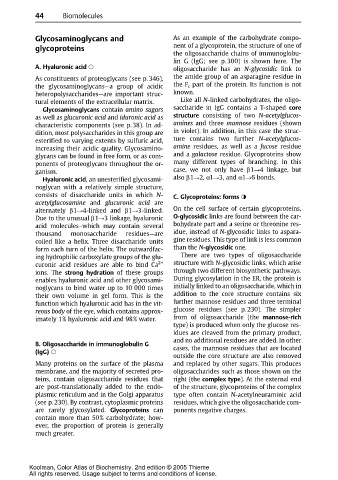
Glycosaminoglycans and As an example of the carbohydrate compo-
glycoproteins nent of a glycoprotein, the structure of one of
the oligosaccharide chains of immunoglobu-
lin G (IgG; see p. 300) is shown here. The
A. Hyaluronic acid oligosaccharide has an N-glycosidic link to
As constituents of proteoglycans (see p. 346), the amide group of an asparagine residue in
the glycosaminoglycans—a group of acidic the F c part of the protein. Its function is not
heteropolysaccharides—are important struc- known.
tural elements of the extracellular matrix. Like all N-linked carbohydrates, the oligo-
Glycosaminoglycans contain amino sugars saccharide in IgG contains a T-shaped core
as well as glucuronic acid and iduronic acid as structure consisting of two N-acetylglucos-
characteristic components (see p. 38). In ad- amines and three mannose residues (shown
dition, most polysaccharides in this group are in violet). In addition, in this case the struc-
esterified to varying extents by sulfuric acid, ture contains two further N-acetylglucos-
increasing their acidic quality. Glycosamino- amine residues, as well as a fucose residue
glycans can be found in free form, or as com- and a galactose residue. Glycoproteins show
ponents of proteoglycans throughout the or- many different types of branching. In this
ganism. case, we not only have β1 4linkage,but
Hyaluronic acid, an unesterified glycosami- also β1 2, α1 3, and α1 6bonds.
noglycan with a relatively simple structure,
consists of disaccharide units in which N- C. Glycoproteins: forms
acetylglucosamine and glucuronic acid are
alternately β1 4-linked and β1 3-linked. On the cell surface of certain glycoproteins,
Due to the unusual β1 3linkage,hyaluronic O-glycosidic links are found between the car-
acid molecules–which may contain several bohydrate part and a serine or threonine res-
thousand monosaccharide residues—are idue, instead of N-glycosidic links to aspara-
coiled like a helix. Three disaccharide units gine residues. This type of link is less common
form each turn of the helix. The outwardfac- than the N-glycosidic one.
ing hydrophilic carboxylate groups of the glu- There are two types of oligosaccharide
curonic acid residues are able to bind Ca 2+ structure with N-glycosidic links, which arise
ions. The strong hydration of these groups through two different biosynthetic pathways.
enables hyaluronic acid and other glycosami- During glycosylation in the ER, the protein is
noglycans to bind water up to 10 000 times initially linked to an oligosaccharide, which in
their own volume in gel form. This is the addition to the core structure contains six
function which hyaluronic acid has in the vit- further mannose residues and three terminal
reous body of the eye, which contains approx- glucoseresidues (seep. 230). Thesimpler
imately 1% hyaluronic acid and 98% water. from of oligosaccharide (the mannose-rich
type) is produced when only the glucose res-
idues are cleaved from the primary product,
and no additional residues are added. In other
B. Oligosaccharide in immunoglobulin G
cases, the mannose residues that are located
(IgG)
outside the core structure are also removed
Many proteins on the surface of the plasma andreplacedby other sugars.Thisproduces
membrane, and the majority of secreted pro- oligosaccharides such as those shown on the
teins, contain oligosaccharide residues that right (the complex type). At the external end
are post-translationally added to the endo- of the structure, glycoproteins of the complex
plasmic reticulum and in the Golgi apparatus type often contain N-acetylneuraminic acid
(see p. 230). By contrast, cytoplasmic proteins residues, which give the oligosaccharide com-
are rarely glycosylated. Glycoproteins can ponents negative charges.
contain more than 50% carbohydrate; how-
ever, the proportion of protein is generally
much greater.
Koolman, Color Atlas of Biochemistry, 2nd edition © 2005 Thieme
All rights reserved. Usage subject to terms and conditions of license.