
Page 95 - Color Atlas of Biochemistry
P. 95
86 Biomolecules
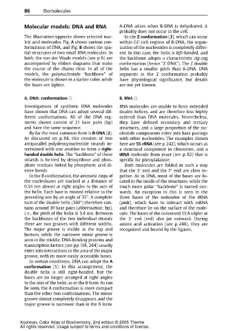
Molecular models: DNA and RNA A-DNA arises when B-DNA is dehydrated. It
probably does not occur in the cell.
The illustration opposite shows selected nuc- In the Z-conformation (3), which can occur
leic acid molecules. Fig. A shows various con- within GC-rich regions of B-DNA, the organ-
formations of DNA, and Fig. B shows the spa- ization of the nucleotides is completely differ-
tial structures of two small RNA molecules. In ent. In this case, the helix is left-handed, and
both, the van der Waals models (see p. 6) are thebackboneadopts a characteristic zig-zag
accompanied by ribbon diagrams that make conformation (hence “Z-DNA”). The Z double
the course of the chains clear. In all of the helix has a smaller pitch than B-DNA. DNA
models, the polynucleotide “backbone” of segments in the Z conformation probably
the molecule is shown in a darker color, while have physiological significance, but details
the bases are lighter. are not yet known.
A. DNA: conformation B. RNA
Investigations of synthetic DNA molecules RNA molecules are unable to form extended
have shown that DNA can adopt several dif- double helices, and are therefore less highly
ferent conformations. All of the DNA seg- ordered than DNA molecules. Nevertheless,
ments shown consist of 21 base pairs (bp) they have defined secondary and tertiary
and have the same sequence. structures, and a large proportion of the nu-
By far the most common form is B-DNA (2). cleotide components enter into base pairings
As discussed on p. 84, this consists of two with other nucleotides. The examples shown
antiparallel polydeoxynucleotide strands in- here are 5S-rRNA (see p. 242), which occurs as
tertwined with one another to form a right- a structural component in ribosomes, and a
handed double helix. The “backbone” of these tRNA molecule from yeast (see p. 82) that is
strandsisformedby deoxyribose and phos- specific for phenylalanine.
phate residues linked by phosphoric acid di- Both molecules are folded in such a way
ester bonds. that the 3 end and the 5 end are close to-
In the B conformation, the aromatic rings of gether. Asin DNA, most of the basesare lo-
the nucleobases are stacked at a distance of cated in the inside of the structures, while the
0.34 nm almost at right angles to the axis of much more polar “backbone” is turned out-
the helix. Each base is rotated relative to the wards. An exception to this is seen in the
preceding one by an angle of 35°. A complete three bases of the anticodon of the tRNA
turn of the double helix (360°) therefore con- (pink), which have to interact with mRNA
tains around 10 base pairs (abbreviation: bp), and therefore lie on the surface of the mole-
i. e., the pitch of the helix is 3.4 nm. Between cule. The bases of the conserved CCA triplet at
thebackbones of thetwo individual strands the 3 end (red) also jut outward. During
there are two grooves with different widths. amino acid activation (see p. 248), they are
The major groove is visible at the top and recognized and bound by the ligases.
bottom, while the narrower minor groove is
seen in the middle. DNA-binding proteins and
transcription factors (see pp.118, 244) usually
enter into interactions in the area of the major
groove, with its more easily accessible bases.
In certain conditions, DNA can adopt the A
conformation (1). In this arrangement, the
double helix is still right-handed, but the
bases are no longer arranged at right angles
to the axis of the helix, as in the B form. As can
be seen, the A conformation is more compact
than the other two conformations. The minor
groove almost completely disappears, and the
major groove is narrower thaninthe B form.
Koolman, Color Atlas of Biochemistry, 2nd edition © 2005 Thieme
All rights reserved. Usage subject to terms and conditions of license.