
Page 207 - Computational Modeling in Biomedical Engineering and Medical Physics
P. 207
196 Computational Modeling in Biomedical Engineering and Medical Physics
and bone resorption and in addition, osteophyte formation alters the bone structures
of the joints. In addition, excessive fibrosis of the fibrous capsule and the inside of the
joint occurs, reducing the function of the normal joint by limiting flexion and exten-
sion. Therefore the biocompatibility of SPION MNPs, their eventual distribution and
elimination are important issue (Shi and Gu, 2008).
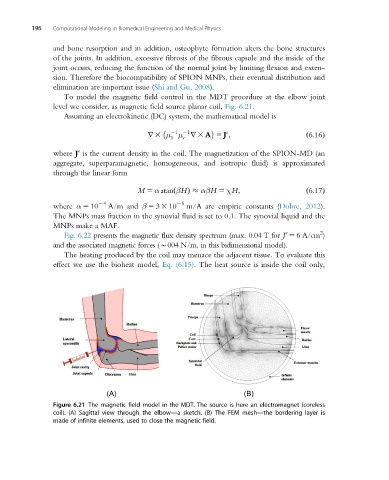
To model the magnetic field control in the MDT procedure at the elbow joint
level we consider, as magnetic field source planar coil, Fig. 6.21.
Assuming an electrokinetic (DC) system, the mathematical model is
1 1 e
r 3 μ μ r 3 A 5 J ;
0 r ð6:16Þ
e
where J is the current density in the coil. The magnetization of the SPION-MD (an
aggregate, superparamagnetic, homogeneous, and isotropic fluid) is approximated
through the linear form
M 5 α atan βHÞ αβH 5 χH; ð6:17Þ
ð
24 25
where α 5 10 A/m and β 5 3 3 10 m/A are empiric constants (Dobre, 2012).
The MNPs mass fraction in the synovial fluid is set to 0.1. The synovial liquid and the
MNPs make a MAF.
2
e
Fig. 6.22 presents the magnetic flux density spectrum (max. 0.04 T for J 5 6A/cm )
and the associated magnetic forces (B004 N/m, in this bidimensional model).
The heating produced by the coil may menace the adjacent tissue. To evaluate this
effect we use the bioheat model, Eq. (6.15). The heat source is inside the coil only,
Figure 6.21 The magnetic field model in the MDT. The source is here an electromagnet (coreless
coil). (A) Sagittal view through the elbow—a sketch. (B) The FEM mesh—the bordering layer is
made of infinite elements, used to close the magnetic field.