
Page 215 - Computational Modeling in Biomedical Engineering and Medical Physics
P. 215
204 Computational Modeling in Biomedical Engineering and Medical Physics
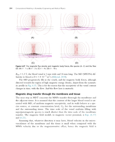
Figure 6.27 The magnetic flux density and magnetic body forces, the species (A C) and the flow
(D). (A) t 5 1 s. (B) t 5 3 s. (C) t 5 4 s. (D) t 5 16 s.
B rem 5 1.3 T, the blood vessel is 1 mm wide and 16 mm long. The MD (SPIONs) dif-
2
fusivity in blood is D 5 1 3 10 29 m /s (Sillerud, 2018).
The MD progressively fills in the vessels, and the magnetic body forces, although
directed towards the region of high magnetic energy density, depart from the symmet-
ric profile in Fig. 6.15. This is for the reason that the susceptivity of the vessel content
changes in time, with the flow. And the flow here is unsteady.
Magnetic drug transfer through the membrane and tissue
The next step in MDT concerns the MNPs transfer thorough the membrane and
the adjacent tissue. It is assumed that the content of the larger blood vessel is sat-
urated with MD, of uniform magnetic susceptivity, and its walls behave as a spe-
cies source, at constant concentration level, C b , for the surrounding membrane
and the surrounding tissue. The time scale of the vessel uniform filling with
superparamagnetic species is much shorter than the time scale of the membrane
transfer. The magnetic field model, in magnetic vector potential, is Eqs. (6.17)
and (6.21).
Assuming that, whatever direction it may have, blood velocity in the micro-
vasculature of the membrane and the tissue is small when compared with the
MNPs velocity due to the magnetomotive effect, hence the magnetic field is