
Page 143 - Computational Retinal Image Analysis
P. 143
1 Introduction 137
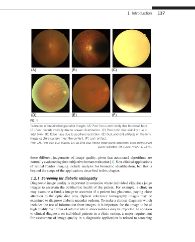
FIG. 1
Examples of impaired/ungradable images. (A) Poor focus and clarity due to overall haze.
(B) Poor macula visibility due to uneven illumination. (C) Poor optic disc visibility due to
total blink. (D) Edge haze due to pupillary restriction. (E) Dust and dirt artifacts on the lens
image capture system (near the center). (F) Lash artifact.
From J.M. Pires Dias, C.M. Oliveira, L.A. da Silva Cruz, Retinal image quality assessment using generic image
quality indicators, Inf. Fusion 19 (2014) 73–90.
these different judgements of image quality, given that automated algorithms are
normally evaluated against subjective human evaluation [1]. Non-clinical applications
of retinal fundus imaging include analysis for biometric identification, but this is
beyond the scope of the applications described in this chapter.
1.2.1 Screening for diabetic retinopathy
Diagnostic image quality is important in scenarios where individual clinicians judge
images to ascertain the ophthalmic health of the patient. For example, a clinician
may examine a fundus image to ascertain if a patient has glaucoma, paying close
attention to the optic disc area. Optical coherence tomography images may be
examined to diagnose diabetic macular oedema. To make a clinical diagnosis which
includes the use of information from images, it is important for the image to be of
high quality over areas of interest where abnormalities may be expected. In addition
to clinical diagnosis on individual patients in a clinic setting, a major requirement
for assessment of image quality in a diagnostic application is related to screening