
Page 17 - Computational Retinal Image Analysis
P. 17
6 CHAPTER 2 Clinical motivation
Subjective impression + Objective measurement
Qualitative assessment +Quantitative assessment
Apparently visible changes + Subtle subclinical changes
On-site diagnosis +Off-site diagnosis
Repetitive work + Automation
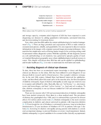
FIG. 1
What values does RIA add to the current medical assessment?
and storage capacity, computer aided diagnosis (CAD) has been expected to assist
diagnosing eye diseases by adding quantitative information, automated detection,
and decision making for treatment choice.
Now, what value does RIA add to the current ophthalmological or medical manage-
ment? (Fig. 1) There are huge potentials and expectations in RIA to make retinal as-
sessment more precise, reliable, and quantifiable. It is also expected to discover unseen
information in the images with computer assessed image processing techniques. Also,
repetitive but simple tasks such as filtering fundus images in the screening setting could
be automated within a diagnostic system. With this capability, off-site diagnosis, known
as a tele-ophthalmology or tele-medicine, has a potential to spread a quality of medi-
cal standard throughout geographic areas where ophthalmological medical resource is
scarce. This chapter will discuss how RIA has and can be applied in ophthalmology
and broader healthcare (Fig. 2) in order to understand the motivation and needs.
1.1 Assisting diagnosis of clinical eye diseases
To date, there has been an accumulated expert knowledge to detect and diagnose
clinical eye diseases on the retina. RIA has been utilized to assist diagnosis of eye
diseases both in research and in clinical practice. One example is diabetic retinopa-
thy. In the mid 19th century, Eduard Jëger had himself improved the ophthalmo-
scope, and described yellowish round spots in the macular area, the first description
of diabetic retinopathy. Signs of diabetic retinopathy at early stage are relatively easy
to recognize and defined by rules. Basically, they are “red lesions,” i.e., microaneu-
rysms and hemorrhages, and hard “yellow lesions,” i.e., hard and soft exudates. To
date, diabetic retinopathy is one eye disease studied for CAD and automated detec-
tion intensively.
There are two reasons why CAD and automated detection of diabetic retinopathy
have been studied extensively. First, there is a clear medical need. The prevalence
of diabetes has quadrupled since 1980, and it is estimated that at present the disease
affects 422 million adults world-wide [3]. Diabetic retinopathy is the most common
complication in diabetes and almost universal in patients with long-term diabetes
[4, 5]. Even though the risk of blindness is estimated to decrease, long-term data have
demonstrated a 25-year incidence of blindness of 9.5% in patients with type 1 dia-
betes [6]. Screening for diabetic retinopathy among patients with diabetes is consid-
ered an important strategy to prevent blindness or severe visual impairment. Indeed,
diabetic retinopathy is a good candidate to be screened at the clinically asymptomatic