
Page 18 - Computational Retinal Image Analysis
P. 18
1 Introduction 7
#1 Assisting
diagnosis of
clinical eye
diseases
#2 Assessing
#5 Structural severity and
signs to
functional classifying
signs Retinal clinical eye
diseases
Image
Analysis
#4 Identifying
retinal #3 Capturing
changes pre-clinical
associated signs of the
with systemic eye diseases
diseases
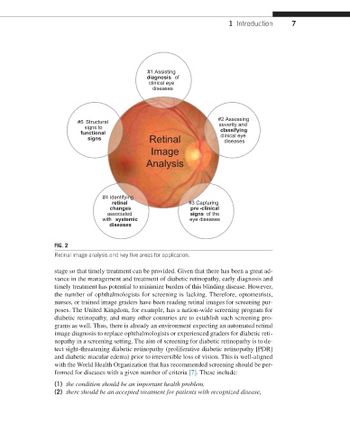
FIG. 2
Retinal image analysis and key five areas for application.
stage so that timely treatment can be provided. Given that there has been a great ad-
vance in the management and treatment of diabetic retinopathy, early diagnosis and
timely treatment has potential to minimize burden of this blinding disease. However,
the number of ophthalmologists for screening is lacking. Therefore, optometrists,
nurses, or trained image graders have been reading retinal images for screening pur-
poses. The United Kingdom, for example, has a nation-wide screening program for
diabetic retinopathy, and many other countries are to establish such screening pro-
grams as well. Thus, there is already an environment expecting an automated retinal
image diagnosis to replace ophthalmologists or experienced graders for diabetic reti-
nopathy in a screening setting. The aim of screening for diabetic retinopathy is to de-
tect sight-threatening diabetic retinopathy (proliferative diabetic retinopathy [PDR]
and diabetic macular edema) prior to irreversible loss of vision. This is well-aligned
with the World Health Organization that has recommended screening should be per-
formed for diseases with a given number of criteria [7]. These include:
(1) the condition should be an important health problem,
(2) there should be an accepted treatment for patients with recognized disease,