
Page 23 - Computational Retinal Image Analysis
P. 23
12 CHAPTER 2 Clinical motivation
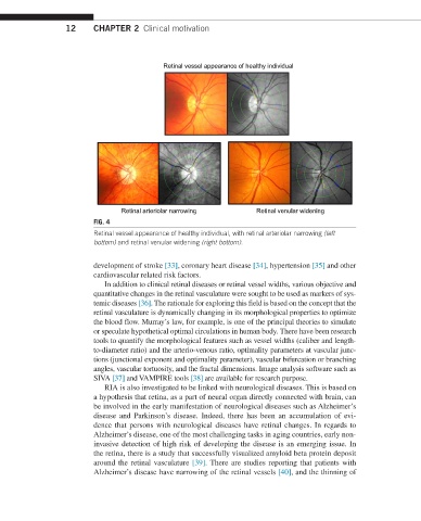
Retinal vessel appearance of healthy individual
Retinal arteriolar narrowing Retinal venular widening
FIG. 4
Retinal vessel appearance of healthy individual, with retinal arteriolar narrowing (left
bottom) and retinal venular widening (right bottom).
development of stroke [33], coronary heart disease [34], hypertension [35] and other
cardiovascular related risk factors.
In addition to clinical retinal diseases or retinal vessel widths, various objective and
quantitative changes in the retinal vasculature were sought to be used as markers of sys-
temic diseases [36]. The rationale for exploring this field is based on the concept that the
retinal vasculature is dynamically changing in its morphological properties to optimize
the blood flow. Murray’s law, for example, is one of the principal theories to simulate
or speculate hypothetical optimal circulations in human body. There have been research
tools to quantify the morphological features such as vessel widths (caliber and length-
to-diameter ratio) and the arterio-venous ratio, optimality parameters at vascular junc-
tions (junctional exponent and optimality parameter), vascular bifurcation or branching
angles, vascular tortuosity, and the fractal dimensions. Image analysis software such as
SIVA [37] and VAMPIRE tools [38] are available for research purpose.
RIA is also investigated to be linked with neurological diseases. This is based on
a hypothesis that retina, as a part of neural organ directly connected with brain, can
be involved in the early manifestation of neurological diseases such as Alzheimer’s
disease and Parkinson’s disease. Indeed, there has been an accumulation of evi-
dence that persons with neurological diseases have retinal changes. In regards to
Alzheimer’s disease, one of the most challenging tasks in aging countries, early non-
invasive detection of high risk of developing the disease is an emerging issue. In
the retina, there is a study that successfully visualized amyloid beta protein deposit
around the retinal vasculature [39]. There are studies reporting that patients with
Alzheimer’s disease have narrowing of the retinal vessels [40], and the thinning of