
Page 24 - Computational Retinal Image Analysis
P. 24
1 Introduction 13
the retina and choroid [41]. There is a newer finding that retinal vessel density is
decreased both in the retina and choroid [42]. Current challenges include whether
there is a specific change or combination of changes that can be used to stratify risk
specific to different diseases [43].
1.5 Structural signs to functional signs
Although it is often the case that a single retinal image is used for assessment, this is
only capturing a static state. Expectations to RIA include an assessment of dynami-
cally changing features based on real-time monitoring or changes over stimulations.
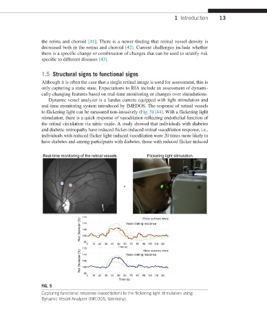
Dynamic vessel analyzer is a fundus camera equipped with light stimulation and
real-time monitoring system introduced by IMEDOS. The response of retinal vessels
to flickering light can be measured non-invasively (Fig. 5) [44]. With a flickering light
stimulation, there is a quick response of vasodilation reflecting endothelial function of
the retinal circulation via nitric oxide. A study showed that individuals with diabetes
and diabetic retinopathy have reduced flicker-induced retinal vasodilation response, i.e.,
individuals with reduced flicker light-induced vasodilation were 20 times more likely to
have diabetes and among participants with diabetes, those with reduced flicker induced
Real-time monitoring of the retinal vessels Flickering light stimulation
+
115 Flicker summary Artery
Rel. Diameter (%) 110 Vaso-dilating response
105
100
95
0 10 20 30 40 50 60 70 80 90 100 110 120
Time (s)
115 Vaso-dilating response
Rel. Diameter (%) 105
Flicker summary Veine
110
100
95
0 10 20 30 40 50 60 70 80 90 100 110 120
Time (s)
FIG. 5
Capturing functional response (vasodilation) to the flickering light stimulation using
Dynamic Vessel Analyzer (IMEDOS, Germany).