
Page 22 - Computational Retinal Image Analysis
P. 22
1 Introduction 11
100
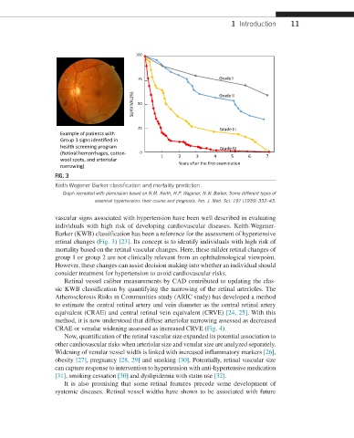
75 Grade I
SURVIVAL(%) 50 Grade II
25 Grade III
Example of patients with
Group 3 signs identified in
health screening program Grade IV
(Retinal hemorrhages, cotton- 0
wool spots, and arteriolar 1 2 3 4 5 6 7
narrowing) Years after the first examination
FIG. 3
Keith Wegener Barker classification and mortality prediction.
Graph recreated with permission based on N.M. Keith, H.P. Wagener, N.W. Barker, Some different types of
essential hypertension: their course and prognosis. Am. J. Med. Sci. 197 (1939) 332–43.
vascular signs associated with hypertension have been well described in evaluating
individuals with high risk of developing cardiovascular diseases. Keith-Wegener-
Barker (KWB) classification has been a reference for the assessment of hypertensive
retinal changes (Fig. 3) [23]. Its concept is to identify individuals with high risk of
mortality based on the retinal vascular changes. Here, these milder retinal changes of
group 1 or group 2 are not clinically relevant from an ophthalmological viewpoint.
However, these changes can assist decision making into whether an individual should
consider treatment for hypertension to avoid cardiovascular risks.
Retinal vessel caliber measurements by CAD contributed to updating the clas-
sic KWB classification by quantifying the narrowing of the retinal arterioles. The
Atherosclerosis Risks in Communities study (ARIC study) has developed a method
to estimate the central retinal artery and vein diameter as the central retinal artery
equivalent (CRAE) and central retinal vein equivalent (CRVE) [24, 25]. With this
method, it is now understood that diffuse arteriolar narrowing assessed as decreased
CRAE or venular widening assessed as increased CRVE (Fig. 4).
Now, quantification of the retinal vascular size expanded its potential association to
other cardiovascular risks when arteriolar size and venular size are analyzed separately.
Widening of venular vessel width is linked with increased inflammatory markers [26],
obesity [27], pregnancy [28, 29] and smoking [30]. Potentially, retinal vascular size
can capture response to intervention to hypertension with anti- hypertensive medication
[31], smoking cessation [30] and dyslipidemia with statin use [32].
It is also promising that some retinal features precede some development of
systemic diseases. Retinal vessel widths have shown to be associated with future