
Page 105 - Academic Press Encyclopedia of Physical Science and Technology 3rd BioTechnology
P. 105
P1: GNB Final Pages
Encyclopedia of Physical Science and Technology EN005F-954 June 15, 2001 20:48
818 Fiber-Optic Chemical Sensors
FIGURE 13 The chemical structure of two dyes that possess
metal-binding crown ethers. The effects of metal ion chelation
will have different photophysical consequences depending on the
location of the interaction. This interaction will cause the dyes to
display unique emission spectra upon metal binding.
approach is not as selective as other methods (discussed
below).
The second ion-sensing approach involves the use of
fluorogenic and chromogenic crown ethers attached to the
fiber tip directly or via an ion-permeable membrane. Flu-
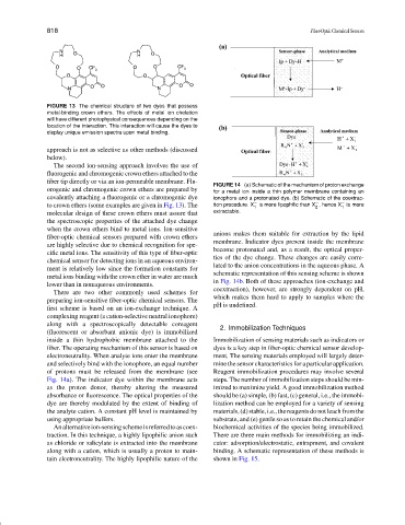
FIGURE 14 (a) Schematic of the mechanism of proton exchange
orogenic and chromogenic crown ethers are prepared by for a metal ion inside a thin polymer membrane containing an
covalently attaching a fluorogenic or a chromogenic dye ionophore and a protonated dye. (b) Schematic of the coextrac-
−
−
−
to crown ethers (some examples are given in Fig. 13). The tion procedure. X is more lipophilic than X , hence X is more
1
1
2
molecular design of these crown ethers must assure that extractable.
the spectroscopic properties of the attached dye change
when the crown ethers bind to metal ions. Ion-sensitive
anions makes them suitable for extraction by the lipid
fiber-optic chemical sensors prepared with crown ethers
membrane. Indicator dyes present inside the membrane
are highly selective due to chemical recognition for spe-
become protonated and, as a result, the optical proper-
cific metal ions. The sensitivity of this type of fiber-optic
ties of the dye change. These changes are easily corre-
chemical sensor for detecting ions in an aqueous environ-
lated to the anion concentrations in the aqueous phase. A
ment is relatively low since the formation constants for
schematic representation of this sensing scheme is shown
metal ions binding with the crown ether in water are much
in Fig. 14b. Both of these approaches (ion-exchange and
lower than in nonaqueous environments.
coextraction), however, are strongly dependent on pH,
There are two other commonly used schemes for
which makes them hard to apply to samples where the
preparing ion-sensitive fiber-optic chemical sensors. The
pH is undefined.
first scheme is based on an ion-exchange technique. A
complexing reagent (a cation-selective neutral ionophore)
along with a spectroscopically detectable coreagent
2. Immobilization Techniques
(fluorescent or absorbant anionic dye) is immobilized
inside a thin hydrophobic membrane attached to the Immobilization of sensing materials such as indicators or
fiber. The operating mechanism of this sensor is based on dyes is a key step in fiber-optic chemical sensor develop-
electroneutrality. When analyte ions enter the membrane ment. The sensing materials employed will largely deter-
and selectively bind with the ionophore, an equal number mine the sensor characteristics for a particular application.
of protons must be released from the membrane (see Reagent immobilization procedures may involve several
Fig. 14a). The indicator dye within the membrane acts steps. The number of immobilization steps should be min-
as the proton donor, thereby altering the measured imized to maximize yield. A good immobilization method
absorbance or fluorescence. The optical properties of the should be (a) simple, (b) fast, (c) general, i.e., the immobi-
dye are thereby modulated by the extent of binding of lization method can be employed for a variety of sensing
the analyte cation. A constant pH level is maintained by materials, (d) stable, i.e., the reagents do not leach from the
using appropriate buffers. substrate, and (e) gentle so as to retain the chemical and/or
Analternativeion-sensingschemeisreferredtoascoex- biochemical activities of the species being immobilized.
traction. In this technique, a highly lipophilic anion such There are three main methods for immobilizing an indi-
as chloride or salicylate is extracted into the membrane cator: adsorption/electrostatic, entrapment, and covalent
along with a cation, which is usually a proton to main- binding. A schematic representation of these methods is
tain electroneutrality. The highly lipophilic nature of the shown in Fig. 15.