
Page 143 - Academic Press Encyclopedia of Physical Science and Technology 3rd Analytical Chemistry
P. 143
P1: GLM/GJK P2: GRB Final Pages
Encyclopedia of Physical Science and Technology En005H-218 June 15, 2001 20:33
Electrophoresis 371
which describes charge and shape; (2) sedimentation co- venient to describe the action as though the middle were a
efficient s, which describes mass, density, and shape; and single section. The middle carried the optical windows on
(3) diffusion coefficient D, which describes shape only. two sides of each channel, and it was usual to monitor the
The common property for these three is shape, which in movementoftheboundarythroughthesewindowsbynon-
turn is contained within the formulation of the frictional invasive optical methods. The experiment was performed
coefficient f [as in Eq. (6)]. Thus, each mobility can be by first introducing the solvent into the cell while all the
related to the other two, provided that the frictional coeffi- sections were connected. Then one would slide the center
cient is identical for the three conditions. Experience has section plus the top section across the bottom, so isolating
shown that this is a valid assumption, provided that exper- the bottom section. The solvent was removed from one
imental conditions are constant during the determinations of the limbs in the ∪-tube and replaced by the solution
(constant composition of solvent, pH, ionic strength, tem- (dialyzed if the experiment involved macroions). The top
perature, and concentration of macroion). Using the ideal section was then moved back, leaving all the sections iso-
relationships for s, D, and u ± [Eqs. (22)–(24)], the rela- lated from the one limb and replaced by solvent before the
tionship for mobility and sedimentation is given in Eq. connection was made to the electrode vessels. The whole
(25): assembly was mounted on a mechanical support, and the
solvent was added in order to fill the electrode vessels and
M(1 − ρ s /ρ m )D
s = (22) top of the cell. Saturated KCl solution was added to the
RT
bottom of each electrode vessel after the electrodes were
D = RT/F (23) inserted. The complete unit was placed in a thermostat-
u ± = Q ± /F (24) ted bath having optical windows for examining the center
section from outside the bath. When it was equilibrated
M(1 − ρ s /ρ m )u ±
s = , (25) for temperature, the middle section was moved across to
Q complete the channel through the ∪-tube. This operation
produced a boundary between solution and solvent at the
where M is the molar mass, ρ s is the solvent density, ρ m
is the macroion weight density, and Q is the charge of two interfaces between top, middle, and bottom sections.
macromolecule calculated from the product of the num- During the experiment, the movement of the boundary was
ber of charges and the charge on an electron. Within the observed by a variety of methods of which the most popu-
maximum allowable experimental conditions that can be larwerschlierenandinterferenceoptics.Thepatternswere
used in sedimentation, this procedure gives velocities that recorded on films, which were eventually measured to cal-
are about 10 times that produced by electrophoretic forces. culate velocities and boundary profiles. Considerable care
This fact explains in part why for many pruposes sedimen- was taken before the experiment began to equalize the col-
tation has proved a more usable preparative technique for umn heights, thus reducing hydrostatic distrubance. The
resolving mixtures than electrophoresis in free solution. electrodes were made from silver wire and coated with
AgCl, so when the electrodes were immersed in the satu-
rated KCl solution at the bottom of the electrode vessels
the major current was transported by Cl . Electrolysis
−
II. FREE-SOLUTION ELECTROPHORESIS
did not occur, provided that the solution under investiga-
+
tion contained K and Cl . The assembly was vertically
−
A. Conventional Procedures
mounted, and the densities of the solutions had to increase
Until the late 1950s electrophoretic experiments were car- from the top to the bottom in order to minimize mechanical
ried out in columns of aqueous solutions. The equipment mixing of the boundaries.
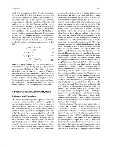
was, in principle, that given in Fig. 1, but, in practice, it Electricalheatingofthesolutionoccurredduringtheex-
consisted of a glass ∪-tube having a square cross-section periment, and the resulting density changes could cause
and constructed from three sections (divided across the convection, which would modify the boundaries and in
channel of the ∪). Each section carried parts of both limbs extreme cases destroy them. To reduce this convection, ex-
of the ∪-tube, and they were designed so that each could periments were normally made at the temperature of maxi-
slide across the others in a plane set normal to the direc- mumdensityofwater(∼277 C)whilethecurrentwaskept
◦
tion of the channels in the ∪-tube. The top section had low by employing organic solutes to buffer the pH when
outlets from the ∪-tube in order to connect the limbs to solutions of polyampholytes were examined. Despite all
separate electrode vessels, while the bottom section was these precautions, considerable experience was required
essentially a connector to complete the bottom part of the in setting up the experiments and interpreting the results.
∪-tube. The middle section was the optical component, This equipment (colloquially called the Tiselius appara-
and it could be divided into two sections, but it is more con- tus) was used extensively by biophysical chemists when