
Page 236 - Academic Press Encyclopedia of Physical Science and Technology 3rd Analytical Chemistry
P. 236
P1: GRB/GWT P2: GPJ/GAX QC: GAE/FYD Final Pages
Encyclopedia of Physical Science and Technology EN008M-395 June 29, 2001 15:52
Magnetic Resonance in Medicine 961
necessary to use special techniques to suppress the very B. Historical Development
strong water signal that tends to overwhelm the signals
It may seem curious that the magnetism of human tissues
from the compounds of interest. Unlike the MRI experi-
can be exploited to develop diagnostic information be-
ment, the information developed from a MRS experiment
cause in normal experience these tissues seem completely
generally does not have a sufficient signal-to-noise ratio
unresponsive to magnetic forces. The explanation is that
(SNR) to permit the display of a highly resolved image
many materials (including water and human tissues), not
showing the distribution in the tissue of the nucleus be-
normally thought of as magnetic, actually possess very
ing studied and of the chemical molecules in which it is
weak magnetic properties that are not evident unless spe-
located. Instead, the data is displayed in the form of a
cial efforts are made to detect them. The magnetic effects
spectrum, which contains peaks associated with various
involved in medical imaging arise from magnetic proper-
compounds containing the nucleus of interest and origi-
ties present in certain atomic nuclei.
nating from a relatively large volume of tissue such as the
The understanding of magnetic properties of materials
liver, brain, or muscle.
has developed synergistically along with other basic phys-
The distinction between imaging and spectroscopy is
ical concepts—particularly atomic structure and quantum
more of a convention based on the mode of excitation and
mechanics—during the twentieth century. In the nine-
display and of the strength of signals detected than it is
teenth century, chemists developed the concept of an
representative of a fundamental distinction in the physical
atom as the irreducible, smallest portion of a chemical
processes involved. Imaging based on water and fat pro-
compound. They established important theoretical con-
tons is less technically demanding than most spectroscopy
cepts such as the periodic table of the chemical elements,
techniques and the results of imaging studies are gener-
and were able to make estimates of atomic size. At this
ally easier to interpret in terms of clinically significant
time, however, there was no understanding of the internal
findings. It is equally true, however, that spectroscopy
structure of the atom, or even a general awareness that such
provides more subtle biochemical, rather than anatomi-
an internal structure existed. Obviously, the concept of nu-
cal, information on the state of the tissues being studied.
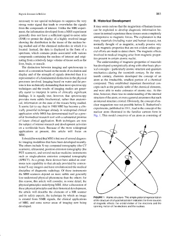
clear magnetism was not possible before E. Rutherford’s
It seems fair to say that in 1988 MRI has become a clin-
experiments, published in 1911, lead to the concept of the
ically powerful technique already in widespread clinical
nuclear atom illustrated in the familiar cartoon form in
application while MRS has demonstrated itself as a pow-
Fig. 1. This model conceives of an atom as consisting of
erful biomedical research tool with a substantial promise
of future clinical application. Both techniques are now
the subject of intense research and development activities
on a worldwide basis. Because of the more widespread
applications at present, this article will focus on
MRI.
ItshouldbenotedthatMRIisbutoneofseveraldiagnos-
tic imaging modalities that have been developed recently.
The others include X-ray computed tomography (the CT
scanners), ultrasound, positron emission tomography (the
PET scanners), and several nuclear medicine instruments
such as single-photon emission computed tomography
(SPECT). As a group, these devices have added an enor-
mous new capability to that already provided by conven-
tional X-ray imagers and have revolutionized the medical
discipline of diagnostic radiology. Of these instruments
the MRI scanners depend on more subtle and generally
less-understood physical phenomena than the others; for
this reason, this article will consider, in some detail, the
physical principles underlying MRI. After a discussion of
these physical principles and their historical development,
the article will describe the elements of a MR scanner
and its safety aspects, the technique by which an image
FIGURE 1 Atomic structure. This simple pictorial representation
is created from NMR signals, the clinical applications
of the structure of a hypothetical atom indicates the three sources
of MRI, and some newer areas of imaging now being of magnetic effects: the orbital motion of the electrons and the
developed. spinning motion of the electrons and the nucleus.