
Page 423 - Academic Press Encyclopedia of Physical Science and Technology 3rd Analytical Chemistry
P. 423
P1: GLQ Final pages
Encyclopedia of Physical Science and Technology EN012C-568 July 26, 2001 15:32
Photoelectron Spectroscopy 73
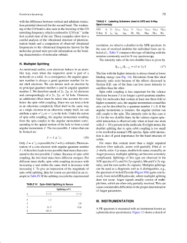
with the difference between vertical and adiabatic ioniza- TABLE V Labeling Schemes Used in XPS and X-Ray
tion potential observed for the second band. The weaken- Spectroscopy
ing of the CO bond is also seen in the lowering of the CO XPS 1s 2s 2p 1/2 2p 3/2 3s 3p 1/2 3p 3/2 3d 3/2 3d 5/2
stretching frequency, which is reduced to 1210 cm −1 in the X-Ray K L I L II L III M I M II M III M IV M V
first excited state of the ion. These examples show how a
careful analysis of the vibrational structure of photoion-
ization bands and a comparison of observed vibrational
resolution, we observe a doublet in the XPE spectrum. In
frequencies to the vibrational frequencies known for the
the case of resolved doublets the individual lines are la-
molecular ground state provide information on the bond-
beled nI J . Table V compares this type of labeling with the
ing characteristics of molecular orbitals.
notation commonly used in X-ray spectroscopy.
The intensity ratio of the two doublet lines is given by
H. Multiplet Splitting
1 = (I + 1)/I (17)
1 /I I− / 2
I I+ / 2
As mentioned earlier, core electrons behave in an atom-
like way, even when the respective atom is part of a The line with the higher intensity is always found at lower
molecule or a solid. As a consequence, the angular quan- binding energy (see Fig. 14). Deviations from this ideal
tum number is always a good quantum number for in- intensity ratio exist because of the effects discussed in
ner shell electrons. We can denote such an electron by Section II.E: one of the lines can lose more intensity to
its principal quantum number n and its angular quantum satellites than the other.
number I. We therefore speak of 2s,2p,or3d electrons Spin–orbit coupling is less important for the valence
and correspondingly of a 2s,2p,or3d hole. However, electrons because I is no longer a good quantum number.
an additional effect must be taken into account for core Only for molecules that contain at least one three-fold or
holes: the spin–orbit coupling. Since we can treat a hole higher symmetry axis, the angular momentum around this
in an otherwise completely filled shell in the same way axis can be described by a quantum number I ≥ 0. If the
as a single electron in an otherwise empty shell, we can angular momentum is nonzero, the angular motion can
1
1
attribute a spin + / 2 or − / 2 to the hole. Under the action still couple to the spin. The intensity ratio in this case is
of spin–orbit coupling, the angular momentum resulting 1:1 for the two doublet lines. In the valence region spin–
from the spin couples to the angular momentum corre- orbit interaction is observed only when at least one atom
sponding to the spatial motion of the hole to form a total with Z > 10 is present in the molecule. For light atoms, the
angular momentum J. The two possible J values that can doublet splitting due to spin–orbit coupling is too small
be formed are to be resolved in normal UPE spectra. Spin–orbit interac-
tion is also of great importance for the band structure of
1
J = I ± / 2 (16)
solids.
1
Only J =+ / 2 is possible for I = 0(s orbitals). Photoion- For states that contain more than a single unpaired
ization of a core electron with angular quantum number electron (free radicals, atoms with partially filled d-or
I > 0 therefore leads to two possible final states that corre- f -shells, nh(n-1)p states, double hole states created by an
spond to the two possible J values. Because of spin–orbit Auger process), multiplet splitting can become extremely
coupling, the two final states have different energies. For complicated. Splittings of this type are observed in the
different inner shells, spin–orbit coupling decreases with XPE spectra of Co and Ni (2p signals), Mn and Cr (3s sig-
increasing n and within the same shell it decreases with nals), and the rare earths (4s signals). Multiplet splittings
increasing I. To give an impression of the magnitude of can be used as a diagnostic tool as it distinguishes, e.g.,
spin–orbit splitting, data for xenon are provided as an ex- the spectrum of nickel(II)oxide (Figure 10b) quite conclu-
ample in Table IV. If the splitting exceeds the experimental sively from nickel(II)hydroxide, where multiplet splitting
does not occur. Auger signals usually consist of multi-
ple lines, which are often only partially resolved. This can
TABLE IV Spin-Orbit Splitting in Xenon
cause considerable difficulties in the proper determination
Splitting (eV)
of Auger parameters.
n I = 1 I = 2
5 1.3
III. INSTRUMENTATION
4 2.0
3 61.5 12.6
A PE spectrum is measured with an instrument known as
2 321.5
a photoelectron spectrometer. Figure 13 shows a sketch of