
Page 141 - Academic Press Encyclopedia of Physical Science and Technology 3rd BioChemistry
P. 141
P1: LDK/GLT P2: GRB Final pages
Encyclopedia of Physical Science and Technology EN013H-614 July 27, 2001 10:29
Protein Folding 181
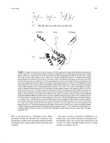
FIGURE 1 Diagram of the levels of protein structure. (A) Amino acids are the basic building blocks (monomers) of
proteins (polymers). All amino acids contain a carboxylic acid group and an amine group connected by a central
carbon called the α-carbon. Each of the 20 common amino acids, designated by a three-letter code, has a unique
side chain (R) that is also bonded to the α-carbon. (B) Through a dehydrolysis reaction, an amide bond is formed
(boxed region) that links the amino group of one amino acid to the carboxylic acid group of the next amino acid.
(C) The primary structure of proteins (1 ) is the linear sequence of amino acids written from the amino-terminal end
◦
(left) to the carboxy-terminal end (right), by convention. Secondary structure of proteins (2 ) is classified into three
◦
major categories. In the α-helix structure, the backbone atoms (amide linkage and α-carbon) coil into a right-handed
1
helical shape (residues 55 to 67 from staphylococcal nuclease are shown). The α-helix is held together through a
series of hydrogen bonds between the amide hydrogen and the carboxyl oxygen of the backbone atoms from amino
acids further up the chain. The side chains (not shown) protrude from the central core structure like the spokes of a
wheel. Another important secondary structure type is the turn (residues 76 to 88 from Staphyloccocal nuclease are
shown). We use the term turn loosely here to represent the regions of proteins that turn corners, thereby allowing
interactions between different and often distant (in terms of primary structure) substructures. The β-sheet structure is
the third common secondary structure type (residues 9 to 12 and 72 to 76 from Staphylococcal nuclease are shown).
It is similar to the α-helical structure in that hydrogen bonds between backbone atoms hold the structure together
and the side chains (not shown) protrude from the structure above and below the plane of the sheet. In contrast to
the α-helix, the β-sheet can be formed from segments of protein that are far apart in the primary sequence. The
tertiary structure (3 ) is the three-dimensional association of secondary structures into a unique and stable final fold.
◦
A ribbon tracing the backbone atoms of Staphylococcal nuclease is shown. 1,2 . The N-terminus of the protein is in
the bottom right and the C-terminus is in the top left of the figure. No side chains are drawn except that of residues
tryptophan 140.
HCl, as will discussed in a subsequent section. These The native structure of proteins is stabilized by in-
perturbants disrupt the intramolecular interactions that tramolecular, noncovalent interactions including hydro-
hold proteins together. One can imagine that the ensemble gen bonding, ionic, and van der Waals interactions, and
of unfolded states could be influenced by the means used covalent cross-links (disulfide bridges between cysteine
to unfold. residues) according to Eq. (4):