
Page 107 - Academic Press Encyclopedia of Physical Science and Technology 3rd Molecular Biology
P. 107
P1: GTQ Final pages
Encyclopedia of Physical Science and Technology EN017F-788 August 3, 2001 16:27
48 Translation of RNA to Protein
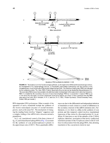
FIGURE 12 Translational control of bacteriophage synthesis. (a) Arrangements of MS2 and f2 bacteriophage cistrons.
(b) Arrangement of Q β bacteriophage cistrons. (c) Replicative intermediate synthesizing new plus strands on the
complementary minus strand copy of the original bacteriophage RNA. The ribosome binding sites (RBS) are indicated
by the numbered arrows. The major RBS (1) binds ribosomes efficiently but can be blocked by ribosomal protein S1.
The secondary RBS (2) becomes available only after translation of at least part of the coat protein cistron by ribosomes.
RBS 3 is masked by the secondary structure of native bacteriophage RNA but is accessible in nascent RNA (c) or
in vitro when the secondary structure is destroyed. Noncoding regions are shown in black upstream from the RBS
sites and at the 3-end. [From Arnstein, H. R. V., and Cox, R. A. (1992). “Protein Synthesis,” Oxford University Press,
Oxford. With permission.]
RNA-dependent DNA polymerase. Other example of the ences are due to the differential and independent initiation
operation of such a frameshift include the synthesis of of translation at each cistron as a result of differences in
the reverse transcriptase enzymes of several retrotrans- the secondary structure of the mRNA initiation sites. Fur-
posons, such as the yeast Ty1. The mechanism of chang- thermore, in vivo there is a delay in the synthesis of coat
ing the reading frame involves “slippery” sequences and protein and this temporal control involves translational
a complex folding of the mRNA into a structure termed a repression of the cistron by ribosomal protein S1. In ad-
pseudoknot. dition, S1 functions as one of the subunits of the f2 RNA
In E. coli, translational control of the three cistrons of replicase; therefore, association of the newly synthesized
Q β ,f2,andrelatedbacteriophageRNAs(Fig.12)accounts translation product of the f2 replicase cistron with S1 will
for the synthesis of coat protein:replicase:A protein in favor its dissociation from the phage RNA, thus allowing
the approximate ratio of 20:5:1. These quantitative differ- translation of the coat protein cistron to start.