
Page 105 - Academic Press Encyclopedia of Physical Science and Technology 3rd Molecular Biology
P. 105
P1: GTQ Final pages
Encyclopedia of Physical Science and Technology EN017F-788 August 3, 2001 16:27
46 Translation of RNA to Protein
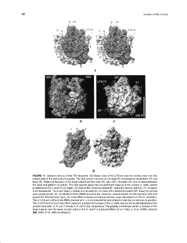
FIGURE 11 Electron density of the 70S ribosome. (A) Stereo view of the 0.78-nm electron density map from the
solvent side of the 30S subunit (purple). The 30S subunit consists of the head (H) connected to the platform (P) and
body (B). Additional features of the small subunit are the neck (N), spur (SP), shoulder (S), and contacts between
the head and platform (a and b). The 50S subunit (gray) has the prominent features of the protein L1 stalk, central
◦
protuberance (CP), and L7/L12 region. (B) View of the ribosome rotated 90 about the vertical, with the L7/L12 region
◦
in the foreground. The A-site finger is marked with an asterisk. (C) View of the ribosome rotated 180 about the vertical
axis compared with (A). (D) Model of three tRNAs bound to the ribosome, viewed toward the 30S interface (left) and
toward the 50S interface (right). All three tRNA surfaces are electron density maps calculated to 0.78-nm resolution.
The 3 -CCA end of the A-site tRNA (marked with ∧) is not modeled for lack of electron density to constrain its position.
The 3 -CCA end of the E-site tRNA (asterisk) is buried at the base of the L1 stalk and cannot be distinguished at the
present resolution. A, P, and E denote A, P, and E site, respectively. The peptidyl transferase center is located on the
large subunit near the lower contact point of the A- and P-site-bound tRNAs. [From Cate, J. et al. (1999). Science
285, 2095–2104. With permission.]