
Page 104 - Academic Press Encyclopedia of Physical Science and Technology 3rd Molecular Biology
P. 104
P1: GTQ Final pages
Encyclopedia of Physical Science and Technology EN017F-788 August 3, 2001 16:27
Translation of RNA to Protein 45
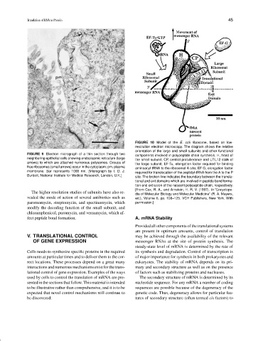
FIGURE 10 Model of the E. coli ribosome, based on low-
resolution electron microscopy. The diagram shows the relative
orientation of the large and small subunits and other functional
FIGURE 9 Electron micrograph of a thin section through two
components involved in polypeptide chain synthesis. H, head of
neighboring epithelial cells showing endoplasmic reticulum (large
the small subunit; CP, central protuberance and L7/L12 stalk of
arrows) to which are attached numerous polysomes. Groups of the large subunit; EF-Tu, elongation factor required for binding
free ribosomes (small arrows) occur in the cytoplasm; pm, plasma aminoacyl-tRNA to the ribosomal A site; EF-G, elongation factor
membrane. Bar represents 1000 nm. [Micrograph by I. D. J. required for translocation of the peptidyl-tRNA from the A to the P
Burdett, National Institute for Medical Research, London, U.K.] site. The broken line indicates the boundary between the transla-
tional and exit domains which are involved in peptide bond forma-
tion and extrusion of the nascent polypeptide chain, respectively.
[From Cox, R. A., and Arnstein, H. R. V. (1997). In “Encyclope-
The higher resolution studies of subunits have also re- dia of Molecular Biology and Molecular Medicine” (R. A. Meyers,
vealed the mode of action of several antibiotics such as ed.), Volume 6, pp. 108–125. VCH Publishers, New York. With
paromomycin, streptomycin, and spectinomycin, which permission.]
modify the decoding function of the small subunit, and
chloramphenicol, puromycin, and vernamycin, which af-
fect peptide bond formation. A. mRNA Stability
Provided all other components of the translational systems
are present in optimum amounts, control of translation
V. TRANSLATIONAL CONTROL may be achieved through the availability of the relevant
OF GENE EXPRESSION messenger RNAs at the site of protein synthesis. The
steady-state level of mRNA is determined by the rate of
Cells needs to synthesize specific proteins in the required its synthesis and degradation. Control of transcription is
amounts at particular times and to deliver them to the cor- of major importance for synthesis in both prokaryotes and
rect locations. These processes depend on a great many eukaryotes. The stability of mRNA depends on its pri-
interactions and numerous mechanisms exist for the trans- mary and secondary structure as well as on the presence
lational control of gene expression. Examples of the ways of factors such as stabilizing proteins and nucleases.
used by cells to control the translation of mRNA are pre- The secondary structure of mRNA is determined by its
sented in the sections that follow. This material is intended nucleotide sequence. For any mRNA a number of coding
to be illustrative rather than comprehensive, and it is to be sequences are possible because of the degeneracy of the
expected that novel control mechanisms will continue to genetic code. Thus, degeneracy allows for particular fea-
be discovered. tures of secondary structure (often termed cis factors) to