
Page 13 - Academic Press Encyclopedia of Physical Science and Technology 3rd Molecular Biology
P. 13
P1: GQQ Revised Pages
Encyclopedia of Physical Science and Technology EN002G-90 May 17, 2001 20:42
Cell Death (Apoptosis) 551
recombinant CAD is recovered in the cytosolic fraction to have chaperone-like activity since the aggregation of
as a CAD/ICAD-L complex. This process is reproduced CAD is suppressed by coexpression with it. Thus ICAD-L
using an in vitro coupled transcription and translation sys- works as a double safeguard against dangerous CAD func-
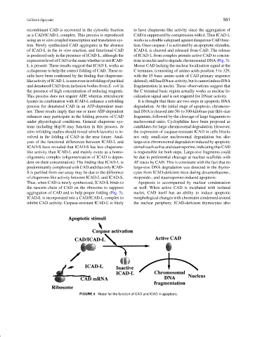
tem. Newly synthesized CAD aggregates in the absence tion. Once caspase-3 is activated by an apoptotic stimulus,
of ICAD-L in the in vitro reaction, and functional CAD ICAD-L is cleaved and released from CAD. The release
is produced only in the presence of ICAD-L, although the of ICAD-L from complex permits active CAD to concen-
expression level of CAD is the same whether or not ICAD- trate in nuclei and to degrade chromosomal DNA (Fig. 5).
L is present. These results suggest that ICAD-L works as Mouse CAD lacking the nuclear localization signal at the
a chaperone to help the correct folding of CAD. These re- C terminus, (consisting of amino acids position 3 to 329,
sults have been confirmed by the finding that chaperone- with the 15 basic amino acids of CAD primary sequence
likeactivityofICAD-Lisseeneveninrefoldingofpurified deleted),stillhasDNaseactivity,butitcannotinduceDNA
and denatured CAD from inclusion bodies from E. coli in fragmentation in nuclei. These observations suggest that
the presence of high concentration of reducing reagents. the C terminal basic region actually works as nuclear lo-
This process does not require ATP, whereas reticulocyte calization signal and is not required for DNase activity.
lysates in combination with ICAD-L enhance a refolding It is thought that there are two steps in apoptotic DNA
process for denatured CAD in an ATP-dependent man- degradation. At the initial stage of apoptosis, chromoso-
ner. These results imply that one or more ATP-dependent mal DNA is cleaved into 50- to 300-kilobase pair (kb)-size
enhancer may participate in the folding process of CAD fragments, followed by the cleavage of large fragments to
under physiological conditions. General chaperone sys- nucleosomal units. Cyclophilins have been proposed as
tems including Hsp70 may function in this process. In candidates for large chromosomal degradation. However,
vitro refolding studies should reveal which factor(s) is in- the expression of caspase-resistant ICAD in cells blocks
volved in the folding of CAD in the near future. Anal- not only small-size nucleosomal degradation but also
yses of the functional differences between ICAD-L and large-size chromosomal degradation induced by apoptotic
ICAD-S have revealed that ICAD-S has less chaperone- stimuli such as Fas and staurosporine, indicating that CAD
like activity than ICAD-L and mainly exists as a homo- is responsible for both steps. Large-size fragments could
oligomeric complex (oligomerization of ICAD is depen- be due to preferential cleavage at nuclear scaffolds with
dent on their concentration). The finding that ICAD-L is AT tracts by CAD. This is consistent with the fact that no
predominantly complexed with CAD and that only ICAD- large-size DNA degradation was detected in the thymo-
S is purified from our assay may be due to the difference cytes from ICAD-deficient mice during dexamethasone-,
of chaperone-like activity between ICAD-L and ICAD-S. etoposide-, and staurosporine-induced apoptosis.
Thus, when CAD is newly synthesized, ICAD-L binds to Apoptosis is accompanied by nuclear condensation
the nascent chain of CAD on the ribosome to suppress as well. When active CAD is incubated with isolated
aggregation of CAD and to help proper folding (Fig. 5). nuclei, CAD itself has an ability to induce apoptotic
ICAD-L is incorporated into a CAD/ICAD-L complex to morphological changes with chromatin condensed around
inhibit CAD activity. Caspase-resistant ICAD-L is likely the nuclear periphery. ICAD-deficient thymocytes also
FIGURE 5 Model for the function of CAD and ICAD in apoptosis.