
Page 21 - Academic Press Encyclopedia of Physical Science and Technology 3rd Molecular Biology
P. 21
P1: GNH Revised Pages
Encyclopedia of Physical Science and Technology EN002G-104 May 17, 2001 20:53
Chromatin Structure and Modification 813
The “linker histone”—histone H1—is so named be-
cause of its physical location in the chromatin fiber (see
following). It is slightly larger than the core histones (ca.
20,000 Da), very rich in lysine, and assumes a very inter-
esting structure shown in Fig. 3, called “the winged helix”
(helix 3 is thought to contact the DNA). Several transcrip-
tional regulators in metazoa are isomorphous to histone
H1, and the implications of this near-congruence for
their physical location within chromatin and mechanisms
whereby they affect gene expression are discussed here.
B. Experimental Evidence for the Histones’
Role in Controlling Gene Expression
Before a discussion of chromatin structure details, it is
helpful to summarize in vivo evidence indicating that his-
tones play a role in gene regulation. Simple a priori con-
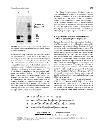
FIGURE 1 The core histones (lane 1), histones H3 and H4, or the
core histones together with the linker histones (lane 3) resolved siderations suffice to realize that histones are required for
on a polyacrylamide gel. cell viability—since chromosome condensation and sub-
sequent segregation during mitosis is contingent on his-
is considerable irony in the brief (ca. 1900–1952) histor- tones, cells would fail to divide in their absence and die,
ical prominence that histones played as the putative ve- and this complicates genetic analysis (as eloquently put
hicles of genetic data: DNA was incorrectly throught to by one molecular biologist, “dead cells don’t tell stories”).
be monotonous in sequence, but histones far exceed the An elegant solution was implemented by M. Grunstein, in
DNA in their invariance, both from nucleosome to nucleo- whose lab a strain of budding yeast, Saccharomyces cere-
some, and between species. Such remarkable resistance to visiae, was engineered for such a study: the promoter of
mutational pressure is particularly striking when one con- the histone H4 gene was replaced with one that could be
siders that—as discussed in the next section—only 75% inactivated with a simple change in growth medium (in
of each histone is actually required to assemble a nucle- this case, the addition of glucose). The gradual depletion
osome core particle. As shown in Fig. 2, all four core of histone H4 from chromatin does not lead to instant cell
histones can be assigned an identical secondary structure: lethality, and thus effects of “genetically dechromatinizing
the COOH-terminal 75% wind into 3 α-helices separated DNA” can be studied.
by two loops (this portion of the histone functions in as- Use of such approaches by Grunstein’s lab, as well as
sembling the histone octamer and in arranging DNA onto of M. Osley, F. Winston, and M. Smith, offered a very
itself), while the structure of the NH 2 -terminal 25% (“the unexpected result: general notions of chromatin as re-
tail”) is not known. The conservation of the primary amino pressive packing material would indicate that most of
acid sequence of the tail is strong evidence for its func- the genome should become spuriously active in the ab-
tional prominence, which is discussed below. sence of chromatin. Experimental observations indicated
FIGURE 2 The core histones. The sequence of the amino terminal tails is indicated. The COOH-terminal domain is
depicted schematically at the bottom.