
Page 23 - Academic Press Encyclopedia of Physical Science and Technology 3rd Molecular Biology
P. 23
P1: GNH Revised Pages
Encyclopedia of Physical Science and Technology EN002G-104 May 17, 2001 20:53
Chromatin Structure and Modification 815
encounters DNA in naked form, random degradation of osome. Two lines of evidence obtained in the mid-1970s
the phosphodiester backbone occurs, and a relatively ho- indicated that the histone proteins lie on the inside of the
mogeneous distribution of DNA sizes is visualized (if nucleosome, while the DNA is somehow exposed on its
the reaction were allowed to proceed longer, DNA would outside surface. M. Noll used the nuclease DNAse I to
be eventually degraded to mononucleotides). In contrast, demonstrate that under appropriate experimental condi-
when cell nuclei are treated with the same nuclease, rather tions, all 146 base pairs of DNA in a single nucleoso-
than degrade the genome in an identical manner, the en- mal particle are cleaved by this nuclease—one cleavage
zyme generates populations of discretely sized DNA frag- was seen to occur every 10−11 base pairs. This suggested
ments that appear to occur in multiples of 180 base pairs. that the nucleic acid is exposed to solution, rather than is
This suggest that in vivo, the DNA is somehow packaged shielded by the histone proteins. More definitive evidence
into “180 base pair installments” such that only the DNA to that effect came from biophysical studies in the labs
stretch between two adjacent 180 base pair “packages” is of B. Richards and C. Crane-Robinson, who used neutron
accessible to the nuclease. At the same time, analysis of scattering to demonstrate that DNA does, indeed, lie on
the hydrodynamic properties of individual DNA–protein the outside of the core histone particle.
particles released by nuclease using analytical centrifuga- In the 20 years that followed, the nucleosome was the
tions, allowed K. van Holde and coworkers to measure its subject of intense investigations. X-ray crystallographic
molecular weight at ca. 180,000 Da. analysis from T. Richmond and A. Klug, and subsequently
Very strong support for the notion of chromatin be- G. Arents and E. Moudrianakis, illuminated the spatial
ing composed of a reiteration of identical subunits came arrangement of its constituents, as did protein-DNA cross-
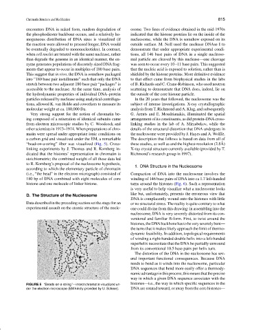
from electron microscopic studies by C. Woodcock and linking studies in the lab of A. Mirzabekov, while the
other scientists in 1973–1974. When preparations of chro- details of the structural distortion that DNA undergoes in
matin were spread under appropriate ionic conditions on the nucleosome were provided by J. Hayes and A. Wolffe.
a carbon grid and visualized under the EM, a remarkable The description that follows is based on data from all of
˚
“bead-on-a-string” fiber was visualized (Fig. 5). Cross- these studies, as well as and the highest-resolution (2.8A)
linking experiments by J. Thomas and R. Kornberg in- X-ray crystal structure currently available (provided by T.
dicated that the histones’ representation in chromatin is Richmond’s research group in 1997).
stoichiometric; the combined weight of all these data led
to R. Kornberg’s proposal of the nucleosome hypothesis,
1. DNA Structure in the Nucleosome
according to which the elementary particle of chromatin
(i.e., “the bead” in the electron micrograph) consisted of Compaction of DNA into the nucleosome involves the
180 bp of DNA combined with eight molecules of core winding of 146 base pairs of DNA into ca 1.7 left-handed
histone and one molecule of linker histone. turns around the histones (Fig. 6). Such a representation
is very useful to help visualize what a nucleosome looks
like but, unfortunately, presents the erroneous view that
D. The Structure of the Nucleosome
DNA is complacently wound onto the histones with little
Data described in the preceding section set the stage for an or no structural stress. The reality is quite contrary to what
experimental assault on the atomic structure of the nucle- one could divine from this drawing: in assembling into the
nucleosome, DNA is very severely distorted from its con-
ventional and familiar B-form. First, to twist around the
histones,theDNAbackbonehastobeveryseverelybent—
the turns that it makes likely approach the limit of thermo-
dynamic feasibility. In addition, topological requirements
of winding a right-handed double helix into a left-handed
superhelix necessitate that the DNA be partially unwound
from its conventional 10.5 base pairs per helix turn.
The distortion of the DNA in the nucleosome has sev-
eral important functional consequences. Because DNA
needs to bend as it winds into the nucleosome, particular
DNA sequences that bend more easily offer a thermody-
namicadvantageinthisprocess;thismeansthattheprecise
way in which a given DNA sequence associates with the
FIGURE 5 “Beads-on-a-string”—insect chromatin visualized un- histones—i.e., the way in which specific sequences in the
der the electron microscope (EM kindly provided by U. Scheer). DNA are rotated toward, or away from the core histones—