
Page 71 - Academic Press Encyclopedia of Physical Science and Technology 3rd Molecular Biology
P. 71
P1: GSS/GLE P2: GPJ Final Pages
Encyclopedia of Physical Science and Technology EN007I-331 July 3, 2001 18:42
682 Immunology—Autoimmunity
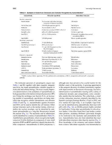
TABLE II Examples of Subcellular Structures and Domains Recognized by Autoantibodies a
Autoantibody Molecular specificity Subcellular structure
Nuclear components
Antichromatin Nucleosomal and subnucleosomal Chromatin
complexes of histones and DNA
Anti-nuclear pore 210-kDa glycoprotein (gp210) Nuclear pore
Antilamin Nuclear lamins A, B, C Nuclear lamina
Anticentromere Centromere proteins (CENP) A, B, C, F Centromere
Anti-p80 coilin p80-coilin (80-kDa protein) Coiled or cajal body
Anti-PIKA p23- to 25-kDa proteins Polymorphic interphase kayrosomal
association (PIKA)
Anti-NuMA 238-kDa protein Mitotic spindle apparatus
Nucleolar components
Antifibrillarin 34-kDa fibrillarin Dense fibrillar component of nucleolus
Anti-RNA polymerase 1 RNA polymerase 1 Fibrillari center of nucleolus
Anti-Pm-Scl 75- and 100-kDa proteins of the Granular component of nucleolus
Pm–Scl complex
Anti-NOR 90 90-kDa doublet of (human) Nucleolar organizer region (NOR)
upstream binding factor (hUBF)
Cytosolic components
Antimitochondria Pyruvate dehydrogenase complex Mitochondria
Antiribosome Ribosomal P proteins (P 0 ,P 1 ,P 2 ) Ribosomes
Anti-Golgi 95- and 160-kDa golgins Golgi apparatus
Antiendosome 180-kDa protein Early endosomes
Antimicrosomal Cytochrome P450 superfamily Microsomes
cANCA Serine proteinase (proteinase 3) Lysosomes
Antimidbody 38-kDa protein Midbody
Anti-centrosome/centriole Pericentrin (48 kDa) Centrosome/centriole
a NuMA, nuclear mitotic apparatus; Pm–Scl, polymyositis–scleroderma; cANCA, cytoplasmic antineutrophil cytoplasmic
antibody.
The molecular spectrum of autoantigenic targets (see although rare, have proven to be a useful marker for the
Tables I and II) together with their exquisite antigenic fibrillar center of the nucleolus. A growing appreciation
specificity has made autoantibodies valuable reagents in of the antigenic diversity of cellular constituents, together
molecular and cellular biology. The most visually impres- with improvements in fluorescent microscopy, has led to
sive demonstration of the usefulness of autoantibodies as identification of autoantibodies reacting with a variety of
biological probes is the indirect immunofluorescence (IIF) subnuclear domains and compartments, some consider-
test. Using this technique (see Section II.A), an increas- ably smaller than the nucleolus. The coiled body, a small
ing number of autoantibody specificities are being iden- circular subnuclear structure originally described by the
tified that recognize cellular substructures and domains Spanish cytologist Santiago Ramon y Cajal in 1903, and
(Table II and Fig. 1). Autoantibodies against chromatin now named the Cajal body, is an example. Cajal bod-
and DNA can be used to identify the cell nucleus. Other ies can be identified using autoantibodies that react with
nuclear structures such as the nuclear lamina, which un- p80 coilin (Fig. 1d), an 80-kDa protein highly enriched
derlies the nuclear envelope, can be identified by anti- in Cajal bodies. Using other autoantibodies in colocaliza-
lamin autoantibodies as a ring-like fluorescence around tion studies, it has been found that Cajal bodies contain
the nucleus (Fig. 1a). The nucleolus and its subdomains other proteins, including fibrillarin (previously thought
can be identified by a variety of autoantibodies (Table II). to be restricted to the nucleolus and prenucleolar bod-
Antifibrillarin autoantibodies, which recognize the highly ies). Autoantibodies have also been identified that react
conserved 34-kDa fibrillarin [a component of some small with subcellular structures other than the nucleus (Fig. 1).
nucleolar RNP (snoRNP) particles], identify the dense fib- Prior knowledge of the existence and relative distribu-
rillar component. Autoantibodies to RNA polymerase I, tions of these subcellular organelles was instrumental in