
Page 75 - Academic Press Encyclopedia of Physical Science and Technology 3rd Molecular Biology
P. 75
P1: GSS/GLE P2: GPJ Final Pages
Encyclopedia of Physical Science and Technology EN007I-331 July 3, 2001 18:42
686 Immunology—Autoimmunity
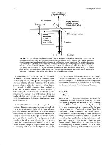
FIGURE 2 Function of filter cube elements in epifluorescence microscopy. The three elements of the filter cube, the
excitation filter, emission filter, and dichroic beam splitting mirror, combine to allow selective use of narrow bandwidths
of radiation in conjunction with specific fluorochromes so that fluorescence can be detected. The excitation filter allows
only certain wavelengths to pass (thick black line), with the dichroic beam splitter reflecting an even narrower bandwidth
toward the specimen. As described by Stokes’ law this excitatory wavelength causes the fluorochrome conjugated
to antibody to emit radiation of a higher wavelength (thick dashed black line), which passes through the dichroic
beam splitter and is transmitted by the emission filter. This radiation is observed as a fluorescent image depicting the
location in the specimen of the antigen bound by the fluorescently labeled antibody.
c. Addition of secondary antibody. The secondary detecting antibody, and the experience of the observer.
(or detecting) antibody (antiserum) is immunoglobulin, Considerable uncertainty in “pattern” recognition can be
usually highly purified, that is specific for the species from avoided through the use of control sera containing defined
which the primary antibody originated. Thus if human autoantibody specificities, such as those available through
serum is being tested for the presence of ANA, then the the Centers for Disease Control, Atlanta, Georgia.
detecting antibody will be anti-human immunoglobulins.
To be useful in immunofluorescence the secondary anti-
body is conjugated to a fluorochrome, most commonly flu- B. ELISA
orescein isothiocyanate (FITC) or rhodamine. However, a
1. History
number of other fluorochrome are also used, including
Alexa 488 and cyanine (Cy3) dyes, which produce more The first report of the use of ELISA (enzyme-linked im-
stable fluorescence. munosorbent assay) to measure antigen specific antibody
was made by Engvall and Perlman in 1972, although
d. Interpretation of results. Under optimal experi- the term ELISA had been used earlier by these work-
mental conditions a serum containing an autoantibody will ers to describe a competitive immunoassay for the quan-
bind to its cognate antigen in the cell or tissue substrate, titative measurement of antigen in solution. Since those
and this bound antibody will in turn be recognized by the early studies ELISA methodology has rapidly expanded
fluorochrome-labeled secondary antibody. When viewed to encompass a variety of techniques for the detection of
through a fluorescence microscope, the emitted fluores- antigen-specific and nonspecific antibodies, soluble and
cence identifies the location of the antigen/autoantibody insoluble antigens, and cellular antigens, indeed any sub-
complex in the cell or tissue substrate. Success in inter- stance which can generate a specific antibody response.
preting the immunofluorescent “pattern” relies on many In autoimmunity the technique has become invaluable as
factors, including the optical properties of the microscope, a screening assay to detect autoantibodies to a variety
the specificity and fluorescent label/protein ratio of the of autoantigens, including proteins and nucleic acids. In