
Page 78 - Academic Press Encyclopedia of Physical Science and Technology 3rd Molecular Biology
P. 78
P1: GSS/GLE P2: GPJ Final Pages
Encyclopedia of Physical Science and Technology EN007I-331 July 3, 2001 18:42
Immunology—Autoimmunity 689
be used. If the task is to screen a group of sera for au-
toantibodies to a particular autoantigen, then care should
be taken to ensure that the level of antigen is sufficient
and that appropriate “negative” controls are used so that
“false-positive” reactions can be eliminated. Considerable
uncertainty in recognition of the appropriate bands for par-
ticular autoantigens can be avoided through the use of con-
trol sera containing relatively monospecific autoantibody
specificities, such as those available through the Centers
for Disease Control, Atlanta, Georgia. Considerable atten-
tion should also be paid to the selection of the secondary
antibody used to detect autoantibody bound to antigen im-
mobilized on nitrocellulose. This reagent should be highly
specific (e.g., affinity purified) and of a high titer. Visual-
ization of the autoantigen/autoantibody complex by sec-
ondary antibody can be achieved in a variety of ways. The
most sensitive methods include conjugation of secondary
antibody with enzymes such as alkaline phosphatase and
horseradish peroxidase. The addition of appropriate sub-
strate allows enzyme-catalyzed colorimetric, fluorescent,
or luminescent reactions that can be readily quantified.
D. Immunoprecipitation
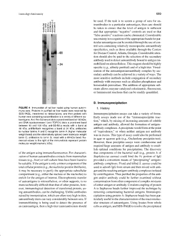
FIGURE 4 Immunoblot of rat liver nuclei using human autoim- 1. History
mune sera. Proteins in purified rat liver nuclei were resolved by
SDS-PAGE, transferred to nitrocellulose, and then probed with Immunoprecipitation assays can take a variety of forms.
human sera containing autoantibodies to a variety of different au- Early assays made use of the “immunoprecipitin reac-
toantigens. Anti-Pm-Scl serum blots a prominent band at 100 kDa; tion,” which, by mixing of increasing amounts of soluble
anti-DNA topoisomerase I (anti-TOPO1) blots a prominent band
between 90 and 100 kDa; anti-SS-B/La reacts with a band at antigen and antibody, allowed the formation of antigen–
48 kDa; antifibrillarin recognizes a band at 34 kDa; antibodies antibody complexes. A precipitate would form at the point
to nuclear lamins A and C recognize lamin A (higher molecular of “equivalence,” or when neither antigen nor antibody
weight band) and the alternatively spliced lower molecular weight was in excess. This type of assay could also be performed
lamin C; antibodies to lamin B 1 react with a 69-kDa band. Nu- in agar or agarose gels (e.g., Oucherlony precipitin test).
merical values to the right of the immunoblots represent protein
molecular weight markers (kDa). However, these precipitin assays were cumbersome and
required large amounts of antigen and antibody to estab-
lish optimal conditions for precipitation. The discovery
of the antigen using immunofluorescence. For characteri- that components of the bacterial wall (e.g., protein A of
zation of human autoantibodies extracts from mammalian Staphyloccus aureus) could bind the Fc portion of IgG
tissues (e.g., liver) or cell culture lines have been found to provided a convenient means of “precipitating” antigen–
be suitable. If the antigen is only a minor component of the antibody complexes. Fixed and killed S. aureus could be
totalcellularprotein(e.g.,thenucleolarproteinfibrillarin), used to adsorb IgG from serum and then mixed with anti-
it may be necessary to purify the appropriate subcellular genandtheresultingantigen–antibodycomplexesisolated
compartment (e.g., either the nucleus or the nucleolus) to by centrifugation. Thus purified the properties of the anti-
enrich for the antigen of interest. Electrophoretic transfer gen and/or antibody could be further examined without
of autoantigens to nitrocellulose, or other supports, is no contaminationfromothercomponentofcomplexmixtures
more technically difficult than that of other proteins, how- of either antigen or antibody. Covalent coupling of protein
ever, immunological detection of transferred protein, us- A to Sepharose beads further improved the technique by
ing autoantibodies, can be challenging. Unlike antibodies removing contaminating bacterial antigens. Immunopre-
raised by immunization, which are often very high in titer, cipitation using protein A–Sepharose beads has been par-
autoantibody titers can vary considerably between sera. If ticularly useful in the characterization of the macromolec-
immunoblotting is being used to detect the presence of ular structure of autoantigens. Using lysates from whole
an (auto)antigen, then a high-titer (auto)antiserum should cells as antigen, and SDS-PAGE to resolve the precipitated