
Page 72 - Academic Press Encyclopedia of Physical Science and Technology 3rd Molecular Biology
P. 72
P1: GSS/GLE P2: GPJ Final Pages
Encyclopedia of Physical Science and Technology EN007I-331 July 3, 2001 18:42
Immunology—Autoimmunity 683
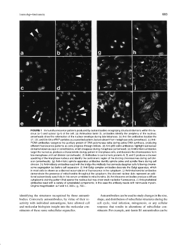
FIGURE 1 Immunofluorescence patterns produced by autoantibodies recognizing structural domains within the nu-
cleus (a–f) and cytosol (g–l) of the cell. (a) Antinuclear lamin B 1 antibodies identify the periphery of the nucleus;
arrowheads show the reformation of the nuclear envelope during late telophase. (b) Anti-Sm antibodies localize the
U1, U2, and U4–U6 snRNP particles as a speckled pattern, but are absent from metaphase cells (arrowhead). (c) Anti-
PCNA antibodies recognize the auxiliary protein of DNA polymerase delta during active DNA synthesis, producing
different fluorescence patterns as cells progress through mitosis. (d) Anti-p80 coilin antibodies highlight subnuclear
domains known as cajal or coiled bodies, which disappear during metaphase (arrowhead). (e) Antifibrillarin antibodies
target the nucleolus, produce a characteristic clumpy pattern in interphase cells, and decorate the chromosomes from
late metaphase until cell division (arrowheads). (f) Antibodies to centromeric proteins A, B, and C produce a discreet
speckling of the interphase nucleus and identify the centromeric region of the dividing chromosomes during cell divi-
sion (arrowheads). (g) Anti-mitotic spindle apparatus antibodies identify spindle poles and spindle fibers during cell
division. (h) Antimidbody antibodies react with the bridge-like midbody that connects daughter cells following chromo-
some segregation but before cell separation. (i) Anti-Golgi complex antibodies decorate the Golgi apparatus, which
in most cells is shown as a discreet accumulation of fluorescence in the cytoplasm. (j) Antimitochondrial antibodies
demonstrate the presence of mitochondria throughout the cytoplasm; the discreet nuclear dots represent an addi-
tional autoantibody specificity in this serum unrelated to mitochondria. (k) Antiribosome antibodies produce a diffuse
cytoplasmic staining pattern that spares the nucleus but may show weak nucleolar fluorescence, (l) Anticytoskeletal
antibodies react with a variety of cytoskeletal components; in this case the antibody reacts with nonmuscle myosin.
Original magnification: a–f and h–l, 350×;g,700×.
identifying the structures recognized by these autoanti- Autoantibodies can be used to study changes in the size,
bodies. Conversely autoantibodies, by virtue of their re- shape, and distribution of subcellular structures during the
activity with individual autoantigens, have allowed cell cell cycle, viral infection, mitogenesis, or any cellular
and molecular biologists insight into the molecular con- response that results in alterations of subcellular con-
stituents of these same subcellular organelles. stituents. For example, anti-lamin B1 autoantiodies can be