
Page 327 - Fiber Fracture
P. 327
FRACTURE OF NATURAL POLYMERIC FIBRES 309
Primary and Secondary Bonds Can Have Direct, Distinguishable, Complementary
Effects on Fibre Mechanical Properties
The charge distribution involved in stabilising a bond can be used to compute the
bond energy, from which the force needed to break the bond can in turn be derived.
Crystallographic information can be used to determine how many such bonds must be
broken per unit area of simple fracture surface. The intrinsic strength of any material
can therefore be calculated from first principles (Kelly and Macmillan, 1986). This
fundamental contribution to strength is often modified at higher length scales. For
example, we have noted in the section ‘A Traditional View of Natural Fibres’ that the
extrinsic properties of conventional textile yams are not related in a simple way to
the intrinsic properties of the constituent polymers; mechanical interactions between
filaments are especially challenging to quantify accurately. In contrast, if we are
concerned with individual filaments that have been produced entirely by self-assembly,
then the properties of the chemical bonds between subunits (at whatever length scale)
will be directly reflected in the properties of the filament.
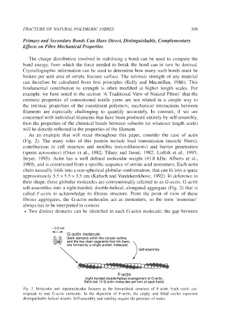
As an example that will recur throughout this paper, consider the case of actin
(Fig. 2). The many roles of this protein include load transmission (muscle fibres),
contributions to cell structure and motility (microfilaments) and barrier penetration
(sperm acrosomes) (Oster et al., 1982; Tilney and InouC, 1982; Lodish et al., 1995;
Stryer, 1995). Actin has a well defined molecular weight (41.8 kDa: Alberts et al.,
1989), and is constructed from a specific sequence of amino acid monomers. Each actin
chain naturally folds into a non-spherical globular conformation, that can fit into a space
approximately 5.5 x 5.5 x 3.5 nm (Kabsch and Vandekerckhove, 1992). In deference to
their shape, these globular molecules are conventionally referred to as G-actin. G-actin
self-assembles into a right-handed, double-helical, elongated aggregate (Fig. 2) that is
called F-actin to acknowledge its fibrous structure. From the point of view of these
fibrous aggregates, the G-actin molecules act as monomers, so the term ‘monomer’
always has to be interpreted in context.
Two distinct domains can be identified in each G-actin molecule; the gap between
- 5.5 nm
U
..<.::... G-actin molecule
..... ,....
..,.,.,.,... (both domains within the circular outline,
.,.\. ..>.,.
and the two chain segments that link them,
$$$ are formed by a single protein molecule)
\ Self-assembly
F-actin
(right-handed double-helical arrangement of G-actin;
there are 13 G-actin molecules per turn of each helix)
Fig. 2. Molecular and supramolecular features in the hierarchical structure of F-actin. Each circle cor-
responds to one G-actin molecule. In the depiction of F-actin, the empty and filled circles represent
distinguishable helical strands. Self-assembly and stability require the presence of water.