Page 177 - Flexible Robotics in Medicine
P. 177
Handheld flexible robot with concentric tubes aiming for intraocular procedures 163

               Similarly for (6.5),
               We can solve for


                                                             x
                                                        21
                                                  θ 5 tan 2                                  (6.10)
                                                             y
               to retain information about each quadrant.
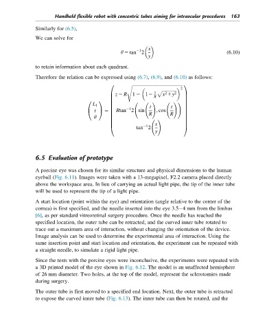
               Therefore the relation can be expressed using (6.7), (6.9), and (6.10) as follows:

                                             0        ffiffiffiffiffiffiffiffiffiffiffiffiffiffiffiffiffiffiffiffiffiffiffiffiffiffiffiffiffiffiffiffiffiffiffiffiffiffiffiffiffiffiffiffiffi 1
                                                    s
                                                                          2

                                                                    2
                                                                       2
                                                z 2 R  1 2 12  1  p ffiffiffiffiffiffiffiffiffiffiffiffiffi
                                                                   x 1y
                                             B                             C
                                                               R
                                             B                             C
                                             B                             C
                                    0    1
                                                            !        !   !
                                      L 1    B                             C
                                             B      21       t        t    C
                                       t   5    Rtan 2 sin      ; cos
                                    @    A   B                             C
                                       θ     B               R        R    C
                                             B
                                                                           C
                                                               !
                                             B                             C
                                                                x
                                             B             21              C
                                                        tan 2
                                             @                             A
                                                                y
               6.5 Evaluation of prototype
               A porcine eye was chosen for its similar structure and physical dimensions to the human
               eyeball (Fig. 6.11). Images were taken with a 13-megapixel, F2.2 camera placed directly
               above the workspace area. In lieu of carrying an actual light pipe, the tip of the inner tube
               will be used to represent the tip of a light pipe.
               A start location (point within the eye) and orientation (angle relative to the center of the
               cornea) is first specified, and the needle inserted into the eye 3.5 4 mm from the limbus
               [6], as per standard vitreoretinal surgery procedure. Once the needle has reached the
               specified location, the outer tube can be retracted, and the curved inner tube rotated to
               trace out a maximum area of interaction, without changing the orientation of the device.
               Image analysis can be used to determine the experimental area of interaction. Using the
               same insertion point and start location and orientation, the experiment can be repeated with
               a straight needle, to simulate a rigid light pipe.

               Since the tests with the porcine eyes were inconclusive, the experiments were repeated with
               a 3D printed model of the eye shown in Fig. 6.12. The model is an unaffected hemisphere
               of 26 mm diameter. Two holes, at the top of the model, represent the sclerotomies made
               during surgery.

               The outer tube is first moved to a specified end location. Next, the outer tube is retracted
               to expose the curved inner tube (Fig. 6.13). The inner tube can then be rotated, and the

