
Page 192 - Flexible Robotics in Medicine
P. 192
Tendon routing and anchoring for cableriven single-t surgical manipulators 179
compression springs only experience a vertical force. Apart from addressing the primary
problem of tendon routing, this technique addresses all the problems experienced from the
2
previous techniques. Since the guide is a piece of 1 mm sheet metal, it does not cause any
adverse hindrance to the flexibility of the spring. Additionally, since a large area of the
guide is in contact with the compression spring body, the guides stay intact with the spring
even after repeated manipulation of the spring. Furthermore, since the guide is sandwiched
between the springs without any protrusion, the overall diameter of the device remains the
same size as the spring diameter. Thus this technique addresses both the design and
functional requirements.
7.4 Integration with surgical tools
After achieving the required actuation method, we now turn toward developing and
integrating the tools. For most of the procedures involving tumor removal, the following
instruments are necessary: a pair of forceps, a cautery device, a suction tube, and an
irrigation channel. Additionally, an endoscopic channel was required to have visual
feedback. The forceps grab onto the tumor for manipulation, while the electrocautery
resects the tumor using heat. The suction tube is then used to isolate and remove the tumor,
water, and blood during the surgical procedure via the use of a vacuum. The endoscope
provides visual feedback by relaying the image of the operating window to a display
monitor. As the overall diameter of the slave device (12 mm) and the diameter of the
individual channels (3 mm) constrains the size of the surgical tools that we can integrate
into the channels, we must make some modifications to the current commercially available
instruments such as the forceps and electrocautery.
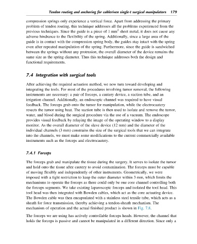
7.4.1 Forceps
The forceps grab and manipulate the tissue during the surgery. It serves to isolate the tumor
and hold onto the tissue after cautery to avoid contamination. The forceps must be capable
of moving flexibly and independently of other instruments. Geometrically, we were
imposed with a tight restriction to keep the outer diameter within 3 mm, which limits the
mechanisms to operate the forceps as there could only be one core channel controlling both
the forceps segments. We take existing laparoscopic forceps and isolated the tool head. This
tool head was then integrated with Bowden cables, which act as the core actuating device.
The Bowden cable was then encapsulated with a stainless steel tensile tube, which acts as a
sheath for force transmission, thereby achieving a tendon-sheath mechanism. The
mechanism of operation and the actual finished product is shown in Fig. 7.8.
The forceps we are using has actively controllable forceps heads. However, the channel that
holds the forceps is passive and cannot be manipulated in a different direction. Since only a