
Page 24 - Flexible Robotics in Medicine
P. 24
6 Chapter 1
(A) (C)
(B) (D)
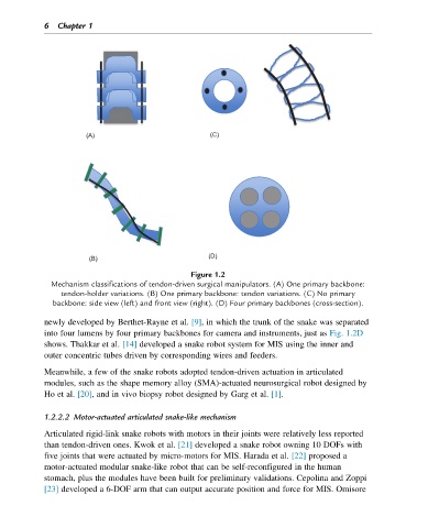
Figure 1.2
Mechanism classifications of tendon-driven surgical manipulators. (A) One primary backbone:
tendon-holder variations. (B) One primary backbone: tendon variations. (C) No primary
backbone: side view (left) and front view (right). (D) Four primary backbones (cross-section).
newly developed by Berthet-Rayne et al. [9], in which the trunk of the snake was separated
into four lumens by four primary backbones for camera and instruments, just as Fig. 1.2D
shows. Thakkar et al. [14] developed a snake robot system for MIS using the inner and
outer concentric tubes driven by corresponding wires and feeders.
Meanwhile, a few of the snake robots adopted tendon-driven actuation in articulated
modules, such as the shape memory alloy (SMA)-actuated neurosurgical robot designed by
Ho et al. [20], and in vivo biopsy robot designed by Garg et al. [1].
1.2.2.2 Motor-actuated articulated snake-like mechanism
Articulated rigid-link snake robots with motors in their joints were relatively less reported
than tendon-driven ones. Kwok et al. [21] developed a snake robot owning 10 DOFs with
five joints that were actuated by micro-motors for MIS. Harada et al. [22] proposed a
motor-actuated modular snake-like robot that can be self-reconfigured in the human
stomach, plus the modules have been built for preliminary validations. Cepolina and Zoppi
[23] developed a 6-DOF arm that can output accurate position and force for MIS. Omisore