
Page 372 - Flexible Robotics in Medicine
P. 372
Robotic transluminal Pan-and-Tilt Scope 365
detect abnormal cell growth or changes in the epithelium. However, this method can only
be performed by a skilled clinician because a careful and precise operation of the
nasopharyngoscope is required. As frequent clinic visits will be mandatory for follow-up
checkups, this method of detection may impose inconvenience to patients and hence,
hinders and delays the detection and treatment of NPC.As a result, treated patients who
have a higher risk of recurrence are deprived of early detection and the capability of
assessing their condition at the ease of their home. If the diagnosis at home is available,
patients will only be required to visit the clinician if any abnormal lesions or bleeding is
observed in the nasal cavity.
Thus there is a need to develop a device that is home-based so that NPC can be regularly
monitored. The device is to be inserted into the nasopharynx through the nostrils. It is desirable
to be easily operated by the patients after specialized training on the use of the device.
Therefore patients would not have to visit the medical centers for consultations periodically.
16.1.2 Approaches addressing the needs
Endoscopic evaluation of the nasopharyngeal region to detect NPC can be accomplished
with both flexible and rigid scopes. Endoscopes began as early as the ancient Greek and
Roman periods. In terms of the evolution of physical characteristics, the differences revolve
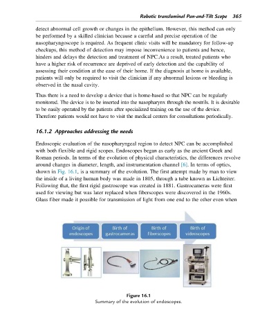
around changes in diameter, length, and instrumentation channel [6]. In terms of optics,
shown in Fig. 16.1, is a summary of the evolution. The first attempt made by man to view
the inside of a living human body was made in 1805, through a tube known as Lichteiter.
Following that, the first rigid gastroscope was created in 1881. Gastrocameras were first
used for viewing but was later replaced when fiberscopes were discovered in the 1960s.
Glass fiber made it possible for transmission of light from one end to the other even when
Figure 16.1
Summary of the evolution of endoscopes.