
Page 419 - Flexible Robotics in Medicine
P. 419
EndoGoose: a flexible and steerable endoscopic forceps 413
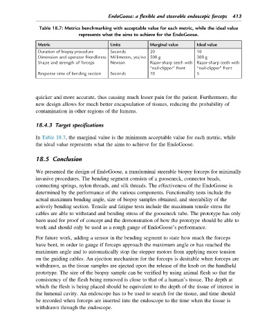
Table 18.7: Metrics benchmarking with acceptable value for each metric, while the ideal value
represents what the aims to achieve for the EndoGoose.
Metric Units Marginal value Ideal value
Duration of biopsy procedure Seconds 20 10
Dimension and operator friendliness Millimeters, yes/no 500 g 300 g
Shape and strength of forceps Newton Razor-sharp teeth with Razor-sharp teeth with
“nail-clipper” front “nail-clipper” front
Response time of bending section Seconds 10 5
quicker and more accurate, thus causing much lesser pain for the patient. Furthermore, the
new design allows for much better encapsulation of tissues, reducing the probability of
contamination in other regions of the lumens.
18.4.3 Target specifications
In Table 18.7, the marginal value is the minimum acceptable value for each metric, while
the ideal value represents what the aims to achieve for the EndoGoose.
18.5 Conclusion
We presented the design of EndoGoose, a transluminal steerable biopsy forceps for minimally
invasive procedures. The bending segment consists of a gooseneck, connector beads,
connecting springs, nylon threads, and silk threads. The effectiveness of the EndoGoose is
determined by the performance of the various components. Functionality tests include the
actual maximum bending angle, size of biopsy samples obtained, and steerability of the
actively bending section. Tensile and fatigue tests include the maximum tensile stress the
cables are able to withstand and bending stress of the gooseneck tube. The prototype has only
been used for proof of concept and the demonstration of how the prototype should be able to
work and should only be used as a rough gauge of EndoGoose’s performance.
For future work, adding a sensor in the bending segment to state how much the forceps
have bent, in order to gauge if forceps approach the maximum angle or has reached the
maximum angle and to automatically stop the stepper motors from applying more tension
on the guiding cables. An ejection mechanism for the forceps is desirable when forceps are
withdrawn, as the tissue samples are ejected upon the release of the knob on the handheld
prototype. The size of the biopsy sample can be verified by using animal flesh so that the
consistency of the flesh being removed is close to that of a human’s tissue. The depth at
which the flesh is being placed should be equivalent to the depth of the tissue of interest in
the lumenal cavity. An endoscope has to be used to search for the tissue, and time should
be recorded when forceps are inserted into the endoscope to the time when the tissue is
withdrawn through the endoscope.