
Page 110 - Fundamentals of Light Microscopy and Electronic Imaging
P. 110
OPTIMIZING THE MICROSCOPE IMAGE 93
(a) (b)
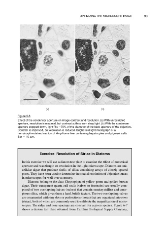
Figure 6-6
Effect of the condenser aperture on image contrast and resolution. (a) With unrestricted
aperture, resolution is maximal, but contrast suffers from stray light. (b) With the condenser
aperture stopped down, light fills 70% of the diameter of the back aperture of the objective.
Contrast is improved, but resolution is reduced. Bright-field light micrograph of a
hematoxylin-stained section of Amphiuma liver containing hepatocytes and pigment cells.
Bar 10 m.
Exercise: Resolution of Striae in Diatoms
In this exercise we will use a diatom test plate to examine the effect of numerical
aperture and wavelength on resolution in the light microscope. Diatoms are uni-
cellular algae that produce shells of silica containing arrays of closely spaced
pores. They have been used to determine the spatial resolution of objective lenses
in microscopes for well over a century.
Diatoms belong to the class Chrysophyta of yellow-green and golden-brown
algae. Their transparent quartz cell walls (valves or frustrules) are usually com-
posed of two overlapping halves (valves) that contain semicrystalline and amor-
phous silica, which gives them a hard, brittle texture. The two overlapping valves
are ornamented with tiny dots or perforations (pores) that are organized into rows
(striae), both of which are commonly used to calibrate the magnification of micro-
scopes. The ridge and pore spacings are constant for a given species. Figure 6-7
shows a diatom test plate obtained from Carolina Biological Supply Company,