
Page 115 - Fundamentals of Light Microscopy and Electronic Imaging
P. 115
98 PHASE CONTRAST MICROSCOPY AND DARK-FIELD MICROSCOPY
5 µm
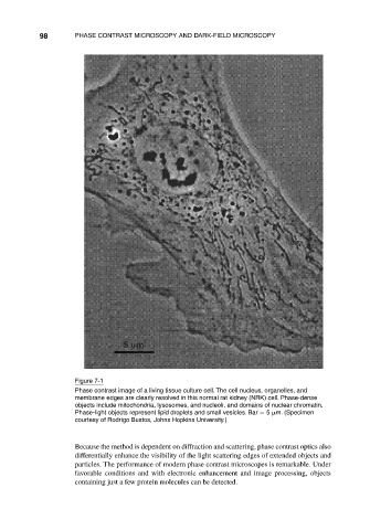
Figure 7-1
Phase contrast image of a living tissue culture cell. The cell nucleus, organelles, and
membrane edges are clearly resolved in this normal rat kidney (NRK) cell. Phase-dense
objects include mitochondria, lysosomes, and nucleoli, and domains of nuclear chromatin.
Phase-light objects represent lipid droplets and small vesicles. Bar 5 m. (Specimen
courtesy of Rodrigo Bustos, Johns Hopkins University.)
Because the method is dependent on diffraction and scattering, phase contrast optics also
differentially enhance the visibility of the light scattering edges of extended objects and
particles. The performance of modern phase contrast microscopes is remarkable. Under
favorable conditions and with electronic enhancement and image processing, objects
containing just a few protein molecules can be detected.