
Page 117 - Fundamentals of Light Microscopy and Electronic Imaging
P. 117
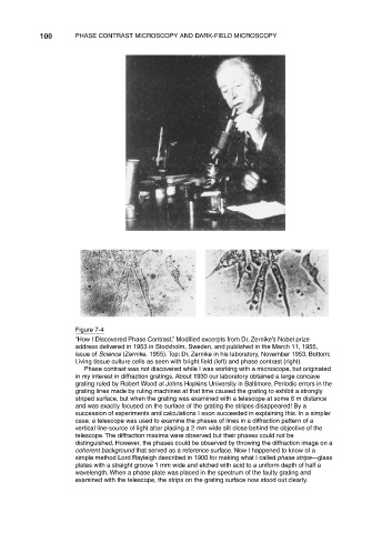
100      PHASE CONTRAST MICROSCOPY AND DARK-FIELD MICROSCOPY


                                Figure 7-4
                                “How I Discovered Phase Contrast.” Modified excerpts from Dr. Zernike’s Nobel prize
                                address delivered in 1953 in Stockholm, Sweden, and published in the March 11, 1955,
                                issue of Science (Zernike, 1955). Top: Dr. Zernike in his laboratory, November 1953. Bottom:
                                Living tissue culture cells as seen with bright field (left) and phase contrast (right).
                                   Phase contrast was not discovered while I was working with a microscope, but originated
                                in my interest in diffraction gratings. About 1930 our laboratory obtained a large concave
                                grating ruled by Robert Wood at Johns Hopkins University in Baltimore. Periodic errors in the
                                grating lines made by ruling machines at that time caused the grating to exhibit a strongly
                                striped surface, but when the grating was examined with a telescope at some 6 m distance
                                and was exactly focused on the surface of the grating the stripes disappeared! By a
                                succession of experiments and calculations I soon succeeded in explaining this. In a simpler
                                case, a telescope was used to examine the phases of lines in a diffraction pattern of a
                                vertical line-source of light after placing a 2 mm wide slit close behind the objective of the
                                telescope. The diffraction maxima were observed but their phases could not be
                                distinguished. However, the phases could be observed by throwing the diffraction image on a
                                coherent background that served as a reference surface. Now I happened to know of a
                                simple method Lord Rayleigh described in 1900 for making what I called phase strips—glass
                                plates with a straight groove 1 mm wide and etched with acid to a uniform depth of half a
                                wavelength. When a phase plate was placed in the spectrum of the faulty grating and
                                examined with the telescope, the strips on the grating surface now stood out clearly.