
Page 121 - Fundamentals of Light Microscopy and Electronic Imaging
P. 121
104 PHASE CONTRAST MICROSCOPY AND DARK-FIELD MICROSCOPY
Image plane
Diffracted
light
Phase plate
Non diffracted
light
Objective
Condenser
Condenser
annulus
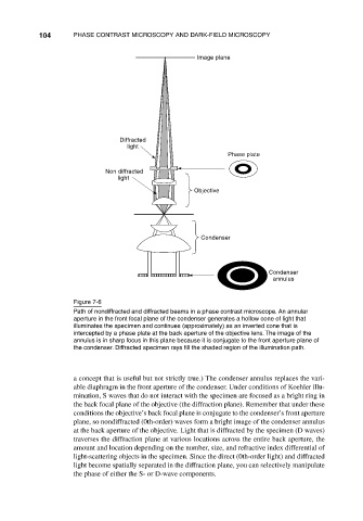
Figure 7-6
Path of nondiffracted and diffracted beams in a phase contrast microscope. An annular
aperture in the front focal plane of the condenser generates a hollow cone of light that
illuminates the specimen and continues (approximately) as an inverted cone that is
intercepted by a phase plate at the back aperture of the objective lens. The image of the
annulus is in sharp focus in this plane because it is conjugate to the front aperture plane of
the condenser. Diffracted specimen rays fill the shaded region of the illumination path.
a concept that is useful but not strictly true.) The condenser annulus replaces the vari-
able diaphragm in the front aperture of the condenser. Under conditions of Koehler illu-
mination, S waves that do not interact with the specimen are focused as a bright ring in
the back focal plane of the objective (the diffraction plane). Remember that under these
conditions the objective’s back focal plane is conjugate to the condenser’s front aperture
plane, so nondiffracted (0th-order) waves form a bright image of the condenser annulus
at the back aperture of the objective. Light that is diffracted by the specimen (D waves)
traverses the diffraction plane at various locations across the entire back aperture, the
amount and location depending on the number, size, and refractive index differential of
light-scattering objects in the specimen. Since the direct (0th-order light) and diffracted
light become spatially separated in the diffraction plane, you can selectively manipulate
the phase of either the S- or D-wave components.