
Page 171 - Fundamentals of Light Microscopy and Electronic Imaging
P. 171
154 DIC MICROSCOPY AND MODULATION CONTRAST MICROSCOPY
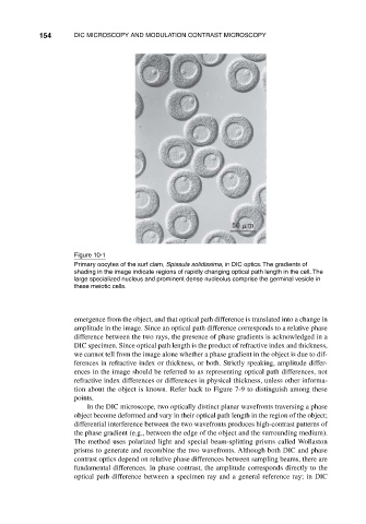
Figure 10-1
Primary oocytes of the surf clam, Spissula solidissima, in DIC optics. The gradients of
shading in the image indicate regions of rapidly changing optical path length in the cell. The
large specialized nucleus and prominent dense nucleolus comprise the germinal vesicle in
these meiotic cells.
emergence from the object, and that optical path difference is translated into a change in
amplitude in the image. Since an optical path difference corresponds to a relative phase
difference between the two rays, the presence of phase gradients is acknowledged in a
DIC specimen. Since optical path length is the product of refractive index and thickness,
we cannot tell from the image alone whether a phase gradient in the object is due to dif-
ferences in refractive index or thickness, or both. Strictly speaking, amplitude differ-
ences in the image should be referred to as representing optical path differences, not
refractive index differences or differences in physical thickness, unless other informa-
tion about the object is known. Refer back to Figure 7-9 to distinguish among these
points.
In the DIC microscope, two optically distinct planar wavefronts traversing a phase
object become deformed and vary in their optical path length in the region of the object;
differential interference between the two wavefronts produces high-contrast patterns of
the phase gradient (e.g., between the edge of the object and the surrounding medium).
The method uses polarized light and special beam-splitting prisms called Wollaston
prisms to generate and recombine the two wavefronts. Although both DIC and phase
contrast optics depend on relative phase differences between sampling beams, there are
fundamental differences. In phase contrast, the amplitude corresponds directly to the
optical path difference between a specimen ray and a general reference ray; in DIC