
Page 173 - Fundamentals of Light Microscopy and Electronic Imaging
P. 173
156 DIC MICROSCOPY AND MODULATION CONTRAST MICROSCOPY
Analyzer
Wollaston II
Objective
Conjugate
interference Object
planes
Condenser
Wollaston I
Polarizer
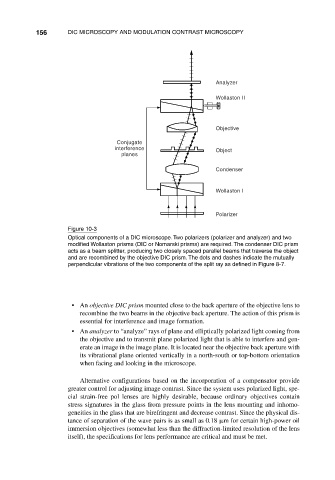
Figure 10-3
Optical components of a DIC microscope. Two polarizers (polarizer and analyzer) and two
modified Wollaston prisms (DIC or Nomarski prisms) are required. The condenser DIC prism
acts as a beam splitter, producing two closely spaced parallel beams that traverse the object
and are recombined by the objective DIC prism. The dots and dashes indicate the mutually
perpendicular vibrations of the two components of the split ray as defined in Figure 8-7.
• An objective DIC prism mounted close to the back aperture of the objective lens to
recombine the two beams in the objective back aperture. The action of this prism is
essential for interference and image formation.
• An analyzer to “analyze” rays of plane and elliptically polarized light coming from
the objective and to transmit plane polarized light that is able to interfere and gen-
erate an image in the image plane. It is located near the objective back aperture with
its vibrational plane oriented vertically in a north-south or top-bottom orientation
when facing and looking in the microscope.
Alternative configurations based on the incorporation of a compensator provide
greater control for adjusting image contrast. Since the system uses polarized light, spe-
cial strain-free pol lenses are highly desirable, because ordinary objectives contain
stress signatures in the glass from pressure points in the lens mounting and inhomo-
geneities in the glass that are birefringent and decrease contrast. Since the physical dis-
tance of separation of the wave pairs is as small as 0.18 m for certain high-power oil
immersion objectives (somewhat less than the diffraction-limited resolution of the lens
itself), the specifications for lens performance are critical and must be met.