
Page 178 - Fundamentals of Light Microscopy and Electronic Imaging
P. 178
THE DIC OPTICAL SYSTEM 161
situation is to decompose the transmitted rays into their corresponding O- and E-wave
components so that we can appreciate the importance of phase displacements between
the waves and the role of the objective DIC prism as a contrasting device. Knowledge of
the action of the objective DIC prism is important, because the operator must adjust the
position of this prism to regulate the amount of optical shadowing and image contrast.
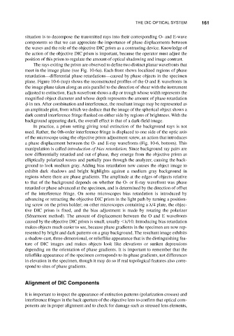
The rays exiting the prism are observed to define two distinct planar wavefronts that
meet in the image plane (see Fig. 10-6a). Each front shows localized regions of phase
retardation—differential phase retardations—caused by phase objects in the specimen
plane. Figure 10-6 (top) shows the reconstructed profiles of the O and E wavefronts in
the image plane taken along an axis parallel to the direction of shear with the instrument
adjusted to extinction. Each wavefront shows a dip or trough whose width represents the
magnified object diameter and whose depth represents the amount of phase retardation
in nm. After combination and interference, the resultant image may be represented as
an amplitude plot, from which we deduce that the image of the spherical object shows a
dark central interference fringe flanked on either side by regions of brightness. With the
background appearing dark, the overall effect is that of a dark-field image.
In practice, a prism setting giving total extinction of the background rays is not
used. Rather, the 0th-order interference fringe is displaced to one side of the optic axis
of the microscope using the objective prism adjustment screw, an action that introduces
a phase displacement between the O- and E-ray wavefronts (Fig. 10-6, bottom). This
manipulation is called introduction of bias retardation. Since background ray pairs are
now differentially retarded and out of phase, they emerge from the objective prism as
elliptically polarized waves and partially pass through the analyzer, causing the back-
ground to look medium gray. Adding bias retardation now causes the object image to
exhibit dark shadows and bright highlights against a medium gray background in
regions where there are phase gradients. The amplitude at the edges of objects relative
to that of the background depends on whether the O- or E-ray wavefront was phase
retarded or phase advanced at the specimen, and is determined by the direction of offset
of the interference fringe. On some microscopes bias retardation is introduced by
advancing or retracting the objective DIC prism in the light path by turning a position-
ing screw on the prism holder; on other microscopes containing a /4 plate, the objec-
tive DIC prism is fixed, and the bias adjustment is made by rotating the polarizer
(Sénarmont method). The amount of displacement between the O and E wavefronts
caused by the objective DIC prism is small, usually /10. Introducing bias retardation
makes objects much easier to see, because phase gradients in the specimen are now rep-
resented by bright and dark patterns on a gray background. The resultant image exhibits
a shadow-cast, three-dimensional, or relieflike appearance that is the distinguishing fea-
ture of DIC images and makes objects look like elevations or sunken depressions
depending on the orientation of phase gradients. It is important to remember that the
relieflike appearance of the specimen corresponds to its phase gradients, not differences
in elevation in the specimen, though it may do so if real topological features also corre-
spond to sites of phase gradients.
Alignment of DIC Components
It is important to inspect the appearance of extinction patterns (polarization crosses) and
interference fringes in the back aperture of the objective lens to confirm that optical com-
ponents are in proper alignment and to check for damage such as stressed lens elements,