
Page 183 - Fundamentals of Light Microscopy and Electronic Imaging
P. 183
166 DIC MICROSCOPY AND MODULATION CONTRAST MICROSCOPY
Image Interpretation
The DIC image has a relieflike quality, exhibiting a shadow-cast effect as if the speci-
men were a three-dimensional surface illuminated by a low-angle light source. It must
be remembered that the shadows and highlights in the shadow-cast image indicate the
sign and slope of phase gradients (gradients in optical path length) in the specimen and
do not necessarily indicate high or low spots. The direction of optical shear is obvious
and is defined by an axis connecting regions having the highest and lowest intensity.
Finally, the direction of the apparent shadow casting reverses for structures with refrac-
tive indices that are lower and higher than the surrounding medium. Thus, dense nuclei,
mitochondria, and lysosomes might have the appearance of raised elevations, while less
dense pinocytotic vesicles and lipid droplets look like sunken depressions. The degree
of contrast and extent of three-dimensionality depend on the amount of bias retardation
between wavefronts imparted by the objective prism. The axis of optical shear cannot be
changed by changing a setting on the microscope. However, the orientation of bright
and dark edges can be reversed 180° by moving the DIC prism to place the optic axis of
the microscope on the other side of the null position of the prism. This has the effect of
reversing the relative phase retardation of the O and E wavefronts. Therefore, the only
way of changing the shear axis relative to the specimen is to rotate the specimen itself.
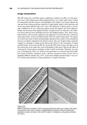
For certain symmetric specimens such as diatoms, specimen rotation can be used to
highlight different features (Fig. 10-8). A precision-rotating specimen stage is very use-
ful in deducing the direction of phase gradients in complex structures.
µ
2m
Figure 10-8
Effect of specimen orientation in DIC microscopy. Since the shear axis is fixed in DIC optics,
the specimen itself must be rotated to highlight different features. Notice the differential
emphasis of pores and striae in the shell of a diatom, Amphipleura, using video-enhanced
DIC optics.