
Page 186 - Fundamentals of Light Microscopy and Electronic Imaging
P. 186
MODULATION CONTRAST MICROSCOPY 169
which uses a related optical system. Like DIC optics, MCM systems produce images
that have a three-dimensional or shadow-cast quality, making objects appear as though
they were illuminated by a low-angle light source (Fig. 10-10). In both MCM and DIC,
brightly illuminated and shadowed edges correspond to optical path gradients (phase
gradients) of opposite slope in the specimen, but unlike DIC, the MCM system does not
require crystalline DIC prisms. Although resolution and detection sensitivity of the
Hoffman MCM system are somewhat reduced compared with DIC, the MCM produces
superior images at lower magnifications, allows optical sectioning of rounded cell spec-
imens, and offers certain advantages over DIC optics, including the ability to examine
cells on birefringent plastic substrates such as cell culture dishes. The Hoffman modu-
lation contrast system is commercially available through Modulation Optics, Inc.,
Greenvale, New York.
Contrast Methods Using Oblique Illumination
Those who test optical surfaces will already be familiar with the essentials of the
schlieren system, which is related to the well-known knife edge test first employed by
Leon Foucault in 1859 for measuring the radius of curvature of a lens surface. Toepler
later used the method to examine variations in the refractive index of a transparent
medium in a sample cell, where inhomogeneities in the medium appear as high contrast
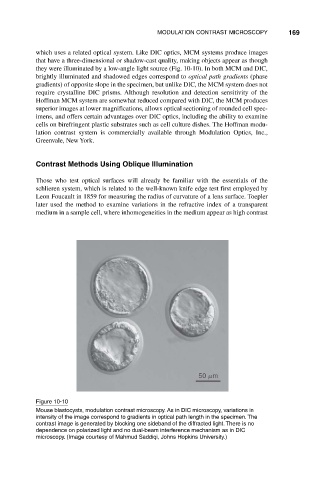
Figure 10-10
Mouse blastocysts, modulation contrast microscopy. As in DIC microscopy, variations in
intensity of the image correspond to gradients in optical path length in the specimen. The
contrast image is generated by blocking one sideband of the diffracted light. There is no
dependence on polarized light and no dual-beam interference mechanism as in DIC
microscopy. (Image courtesy of Mahmud Saddiqi, Johns Hopkins University.)