
Page 185 - Fundamentals of Light Microscopy and Electronic Imaging
P. 185
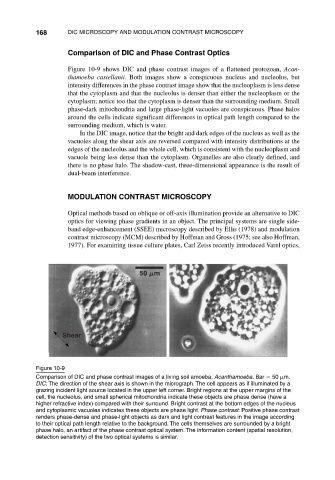
168      DIC MICROSCOPY AND MODULATION CONTRAST MICROSCOPY

                                Comparison of DIC and Phase Contrast Optics

                                Figure 10-9 shows DIC and phase contrast images of a flattened protozoan, Acan-
                                thamoeba castellanii. Both images show a conspicuous nucleus and nucleolus, but
                                intensity differences in the phase contrast image show that the nucleoplasm is less dense
                                that the cytoplasm and that the nucleolus is denser than either the nucleoplasm or the
                                cytoplasm; notice too that the cytoplasm is denser than the surrounding medium. Small
                                phase-dark mitochondria and large phase-light vacuoles are conspicuous. Phase halos
                                around the cells indicate significant differences in optical path length compared to the
                                surrounding medium, which is water.
                                    In the DIC image, notice that the bright and dark edges of the nucleus as well as the
                                vacuoles along the shear axis are reversed compared with intensity distributions at the
                                edges of the nucleolus and the whole cell, which is consistent with the nucleoplasm and
                                vacuole being less dense than the cytoplasm. Organelles are also clearly defined, and
                                there is no phase halo. The shadow-cast, three-dimensional appearance is the result of
                                dual-beam interference.


                                MODULATION CONTRAST MICROSCOPY

                                Optical methods based on oblique or off-axis illumination provide an alternative to DIC
                                optics for viewing phase gradients in an object. The principal systems are single side-
                                band edge-enhancement (SSEE) microscopy described by Ellis (1978) and modulation
                                contrast microscopy (MCM) described by Hoffman and Gross (1975; see also Hoffman,
                                1977). For examining tissue culture plates, Carl Zeiss recently introduced Varel optics,


                               Shear


                       Figure 10-9
                       Comparison of DIC and phase contrast images of a living soil amoeba, Acanthamoeba. Bar   50  m.
                       DIC: The direction of the shear axis is shown in the micrograph. The cell appears as if illuminated by a
                       grazing incident light source located in the upper left corner. Bright regions at the upper margins of the
                       cell, the nucleolus, and small spherical mitochondria indicate these objects are phase dense (have a
                       higher refractive index) compared with their surround. Bright contrast at the bottom edges of the nucleus
                       and cytoplasmic vacuoles indicates these objects are phase light. Phase contrast: Positive phase contrast
                       renders phase-dense and phase-light objects as dark and light contrast features in the image according
                       to their optical path length relative to the background. The cells themselves are surrounded by a bright
                       phase halo, an artifact of the phase contrast optical system. The information content (spatial resolution,
                       detection sensitivity) of the two optical systems is similar.