
Page 187 - Fundamentals of Light Microscopy and Electronic Imaging
P. 187
170 DIC MICROSCOPY AND MODULATION CONTRAST MICROSCOPY
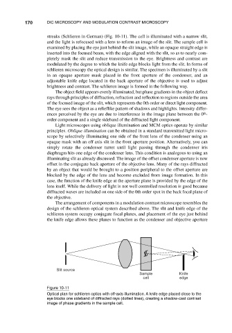
streaks (Schlieren in German) (Fig. 10-11). The cell is illuminated with a narrow slit,
and the light is refocused with a lens to reform an image of the slit. The sample cell is
examined by placing the eye just behind the slit image, while an opaque straight edge is
inserted into the focused beam, with the edge aligned with the slit, so as to nearly com-
pletely mask the slit and reduce transmission to the eye. Brightness and contrast are
modulated by the degree to which the knife edge blocks light from the slit. In forms of
schlieren microscopy the optical design is similar. The specimen is illuminated by a slit
in an opaque aperture mask placed in the front aperture of the condenser, and an
adjustable knife edge located in the back aperture of the objective is used to adjust
brightness and contrast. The schlieren image is formed in the following way.
The object field appears evenly illuminated, but phase gradients in the object deflect
rays through principles of diffraction, refraction and reflection to regions outside the area
of the focused image of the slit, which represents the 0th order or direct light component.
The eye sees the object as a relieflike pattern of shadows and highlights. Intensity differ-
th
ences perceived by the eye are due to interference in the image plane between the 0 -
order component and a single sideband of the diffracted light component.
Light microscopes using oblique illumination and MCM optics operate by similar
principles. Oblique illumination can be obtained in a standard transmitted light micro-
scope by selectively illuminating one side of the front lens of the condenser using an
opaque mask with an off axis slit in the front aperture position. Alternatively, you can
simply rotate the condenser turret until light passing through the condenser iris
diaphragm hits one edge of the condenser lens. This condition is analogous to using an
illuminating slit as already discussed. The image of the offset condenser aperture is now
offset in the conjugate back aperture of the objective lens. Many of the rays diffracted
by an object that would be brought to a position peripheral to the offset aperture are
blocked by the edge of the lens and become excluded from image formation. In this
case, the function of the knife edge at the aperture plane is provided by the edge of the
lens itself. While the delivery of light is not well controlled resolution is good because
diffracted waves are included on one side of the 0th order spot in the back focal plane of
the objective.
The arrangement of components in a modulation contrast microscope resembles the
design of the schlieren optical system described above. The slit and knife edge of the
schlieren system occupy conjugate focal planes, and placement of the eye just behind
the knife edge allows these planes to function as the condenser and objective aperture
Slit source
Sample Knife
cell edge
Figure 10-11
Optical plan for schlieren optics with off-axis illumination. A knife edge placed close to the
eye blocks one sideband of diffracted rays (dotted lines), creating a shadow-cast contrast
image of phase gradients in the sample cell.