
Page 188 - Fundamentals of Light Microscopy and Electronic Imaging
P. 188
MODULATION CONTRAST MICROSCOPY 171
planes in a microscope. The object and retina define two conjugate field planes of the
system. These features are modified in Hoffman modulation contrast optics as shown in
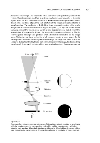
Figure 10-12. An off-axis slit of some width is mounted in the front aperture of the con-
denser, while the knife edge at the back aperture of the objective is represented by a
modulator plate. The modulator is divided into three asymmetric regions: (1) a nearly
opaque section of a circle at the extreme edge of the plate, (2) an adjacent semidarkened
rectangle giving 15% transmission, and (3) a large transparent zone that allows 100%
transmission. When properly aligned, the image of the condenser slit exactly fills the
semitransparent rectangle and produces even, attenuated illumination in the image
plane. Sliding the modulator to the right or left exposes a greater or lesser area of the slit
and brightens or darkens the background in the image. This right-left shear axis is the
same axis that defines the bright and dark contrast regions in the image, but details along
a north-south diameter through the object have minimal contrast. To examine contrast
Bright Dark
Image
Modulator
plate
Objective
Specimen
Condenser
Slit aperture
Figure 10-12
Equipment for modulation contrast microscopy. Oblique illumination is provided by an off-axis
slit in the condenser aperture. A modulator plate with matching complementary slit in the
objective back aperture differentially blocks one sideband of diffracted light. Movement of the
plate modulates the transmission of 0th-order light, allowing for regulation of image contrast.